HUVEC Cell Culture: Human Umbilical Vein Endothelial Cells for Vascular Research
HUVECs (Human Umbilical Vein Endothelial Cells) are primary human endothelial cells isolated from the umbilical cord vein. They are considered the gold standard model for vascular and endothelial research and are widely used in studies of angiogenesis, endothelial function, and cardiovascular biology. HUVECs provide a robust and reproducible platform for investigating human vascular physiology and disease mechanisms.
Key Research Applications of HUVECs
HUVECs are versatile and widely applied in the following areas:
-
- Angiogenesis and vasculogenesis – modeling new blood vessel formation in 2D and 3D systems
- Endothelial function and vascular biology – including endothelial dysfunction studies and procoagulation activity assays
- Cancer research – studying tumor–endothelium interactions
- Wound healing and tissue regeneration – assessing endothelial contribution to repair processes
- Cardiovascular disease studies – such as atherosclerosis and other vascular disorders
Essential Products and Reagents for reliable HUVEC Culture
To ensure reproducible results, high-quality products and careful experimental design are critical:
- HUVECs:
- HUVEC Medium – Endothelial Cell (EC) Culture Media for HUVEC and other EC Types:
- Extracellular Matrices and Synthetic Coatings:
- Collagen I (PureCol, RatCol or VitroCol) – Standard matrix for 2D monolayer culture and 3D HUVEC models, including 3D sprouting and angiogenesis assays, supporting cell adhesion, migration, and proliferation.
- Collagen IV – Mimics the basal membrane and supports physiological endothelial cell structure and differentiation.
- Fibronectin – Promotes migration, angiogenesis, and network formation in wound healing or tissue regeneration assays.
- Vitronectin – Supports adhesion in serum-reduced or defined medium conditions and contains the RGD motiv for integrin-mediated cell attachment.
- Poly-D-Lysine (PDL) and Poly-L-Lysine (PLL) – Synthetic coatings often used in transwell or permeable support systems to improve cell adhesion.
- PEG-based coatings – Biologically inert surfaces for 2D or 3D culture and microfluidic systems; can be functionalized with ligands for specific cell adhesion.
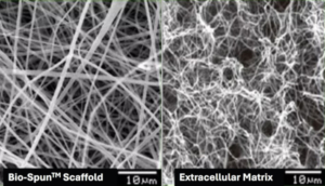
- Scaffolds:
- Bio-Spun™ PU or Bio-SpunTM PET, both applicable for use with smooth muscle cells in arterial model systems.
- Additional critical reagents:
- Subculture Reagents for gentle and reliable passaging of cells.
- High-quality Coagulation Proteins for procoagulation activity assays.
- Carefully selected Cytokines to ensure consistent assay outcomes.
Contact us today to discuss how our HUVECs, defined media, and optimized coatings can help you achieve reproducible, physiologically relevant results.