
Collagen
Structure and Function of Type I Collagen
Collagen type I is a major structural component of skin, bone, tendon, and other fibrous connective tissues, and differs from other collagens by its low lysine hydroxylation and low carbohydrate composition. Although differnt types of collagen have been identified, all are composed of molecules containing three polypeptide chains arranged in a triple helical conformation. Slight differences in the primary structure (amino acid sequence) establish differences between the types.
The amino acid sequence of the primary structure is mainly a repeating motif with glycine in every third position with proline or 4-hydroxyproline frequently preceding the glycine residue. Type I collagen is a heterotrimer composed of two α1(I) chains and one α2(I) chain, which spontaneously form a triple helix scaffold at neutral pH and 37 degrees.
Role of Type I Collagen in Cellular Function
Control of cell growth, differentiation, and apoptosis in multicellular organisms is dependent on adhesion of cells to the extracellular matrix (ECM). Given that Type I collagen is an abundant component of the ECM, cells cultured in three-dimensional (3D) collagen gels simulate the in vivo cell environment better than traditional 2D systems. This has been shown for a number of cell types including cardiac and corneal fibroblasts, hepatic stellate cells (HSCs), and neuroblastoma cells. Several diseases can affect the mechanical properties of the ECM while other disease states may be caused by changes in the density or rigidity of the ECM.
Utilization of Collagen Gels in Cell Culture
Since Type I collagen is a key determinant of tensile properties of the ECM, 3D collagen gels are useful in studies of mechano-transduction, cell signaling involving the transformation of mechanical signals into biochemical signals. 3D gels allow for the study of the effects of the mechanical properties of the ECM, such as density and rigidity, on cell development, migration, and morphology. Unlike 2D systems, 3D environments allow cell extensions to simultaneously interact with integrins on all cell surfaces, resulting in the activation of specific signaling pathways. Gel stiffness or rigidity also affects cell migration differently in 3D versus 2D environments. Furthermore, integrin-independent mechanical interactions resulting from the entanglement of matrix fibrils with cell extensions are possible in 3D systems, but not in 2D systems where the cells are attached to a flat surface.
Cell Surface Receptors for Collagen Subtypes
Different collagen subtypes are recognized by a structurally and functionally diverse group of cell surface receptors, which recognize the collagen triple helix. The best-known collagen receptors are the integrins α1β1 and α2β1. α1β1 is the major integrin on smooth muscle cells, while α2β1 is the major form on epithelial cells and platelets. Both forms are expressed on many cell types including fibroblasts, endothelial cells, osteoblasts, chondrocytes, and lymphocytes. Some cell types may also express other collagen receptors such as glycoprotein VI (GPVI), which mediates both adhesion and signaling in platelets. Other collagen receptors include discoidin domain receptors, leukocyte-associated IG-like receptor-1, and members of the mannose receptor family.
Atelocollagen versus Telocollagen
Telocollagen (Extraction method: acid) and atelocollagen (pepsin treatment) are two forms of collagen that differ in their degree of chemical modification. Telocollagen is the native form of collagen found in the body, containing telopeptides, which are non-helical regions at both ends of the collagen molecule. These telopeptides play a role in cross-linking collagen fibers and stabilizing the collagen structure.
Atelocollagen is a modified form of collagen where the telopeptides have been removed. This removal of telopeptides makes atelocollagen more soluble and less immunogenic compared to telocollagen. Atelocollagen is often used in biomedical applications, such as tissue engineering, wound healing, and cosmetic procedures, due to its enhanced biocompatibility and ability to integrate more effectively with host tissues.
Our Atelocollagens are: PureCol, Nutragen, FibriCol, and VitroCol
Our Telocollagens are: TeloCol-3, TeloCol-6, TeloCol-10, and RatCol
Available Collagen Types for Research
In addition, to the Type I collagen (derived from bovine), the following species are currently available:
VitroCol (human), and RatCol (rat).
In the last decades additional collagen types were analyzed and became commercial available:
Collagen type III (human)
Collagen type IV (human)
-

PureCol®, Bovine Collagen
Cat.-Nr: 5005-100ML
PureCol® collagen (bovine collagen, atelocollagen) is known as the standard of all collagens for purity (>99.9% collagen content), functionality,... Read More
-
Collagen Hybridizing Peptide - 5-FAM Conjugate
Cat.-Nr: 5264-60UG
The Collagen Hybridizing Peptide (CHP) is a synthetic peptide that can specifically bind to denatured collagen strands through hydrogen bonding, both... Read More
-
Collagen Hybridizing Peptide - Biotin Conjugate
Cat.-Nr: 5265-60UG
The Collagen Hybridizing Peptide (CHP) is a synthetic peptide that can specifically bind to denatured collagen strands through hydrogen bonding, both... Read More
-
Collagen Hybridizing Peptides - CY3 Conjugate
Cat.-Nr: 5276-60UG
The Collagen Hybridizing Peptide (CHP) is a synthetic peptide that can specifically bind to denatured collagen strands through hydrogen bonding, both... Read More
-
Collagen, bovine, lyophilized - CL (10g)
Cat.-Nr: LS001656
Type I collagen prepared by the method of Einbinder and Schubert, J. Biol. Chem., 188, 335 (1951). Supplied as a shredded, lyophilized, insoluble... Read More
-
Collagen, bovine, lyophilized - CL (1g)
Cat.-Nr: LS001654
Type I collagen prepared by the method of Einbinder and Schubert, J. Biol. Chem., 188, 335 (1951). Supplied as a shredded, lyophilized, insoluble... Read More
-
Collagen, bovine, lyophilized - CL (5g)
Cat.-Nr: LS001652
Type I collagen prepared by the method of Einbinder and Schubert, J. Biol. Chem., 188, 335 (1951). Supplied as a shredded, lyophilized, insoluble... Read More
-

Collagenase, Powder
Cat.-Nr: 5030-50MG
This Collagenase product is provided as a lyophilized, sterile powder in a 50 mg package size. It has been tested specifically and shown to... Read More
-

FibriCol ™, Bovine Collagen Solution, 10 mg/ml
Cat.-Nr: 5133-20ML
FibriCol® collagen (bovine collagen, atelocollagen) is known as the standard of all collagens for purity (>99% collagen content), functionality,... Read More
-
High Concentration Rat Tail Collagen 10 mg/mL
Cat.-Nr: HCRTC-100MG
RatCol® High Concentration Type I Acid Soluble Rat Tail Collagen (telocollagen) contains 100 mg at a concentration of approximately 10 mg/mL in a... Read More
-

Human Collagen Solution Type III
Cat.-Nr: 5021-10MG
This Type III Collagen product is isolated from human placenta and is purified using a multi-step process with approximately 85% Type III collagen... Read More
-

Human Collagen Type III Powder
Cat.-Nr: 5019-10MG
This Type III Collagen product is isolated from human placenta and is purified using a multi-step process with approximately 85% Type III collagen... Read More
-
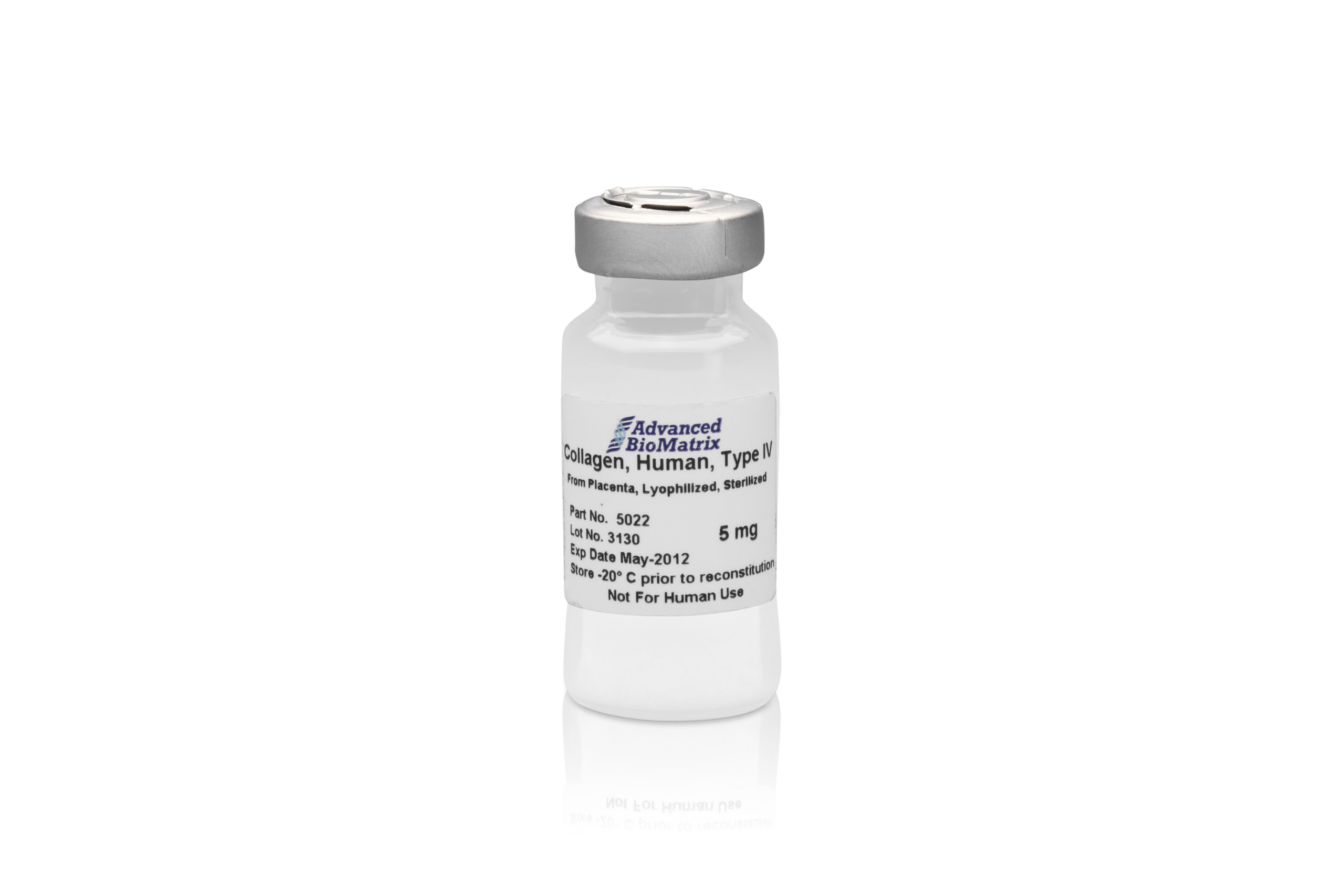
Human Collagen Type IV lyoph.
Cat.-Nr: 5022-5MG
This Type IV Collagen is isolated from human placenta and is purified using a multi-step process. The product is supplied as a sterile, lyophilized... Read More
-

Human Collagen Type IV Powder
Cat.-Nr: 5016-5MG
This Type IV Collagen is isolated from human placenta and is purified using a multi-step process. The product is supplied as a non-sterile powder... Read More
-

Hydrochloric Acid, Solution
Cat.-Nr: 5077-100ML
Hydrochloric Acid (HCl) 0.01M is formulated and prepared to use in conjunction with other products provided by Advanced BioMatrix. HCl is provided... Read More
-

Lifeink® 200; Type I Collagen Bioink
Cat.-Nr: 5278-5ML
For printing with Lifeink® 200, we highly recommend using the FRESH printing technique and printing at 2-8°C. Three dimensional (3D) gels allow for... Read More
-
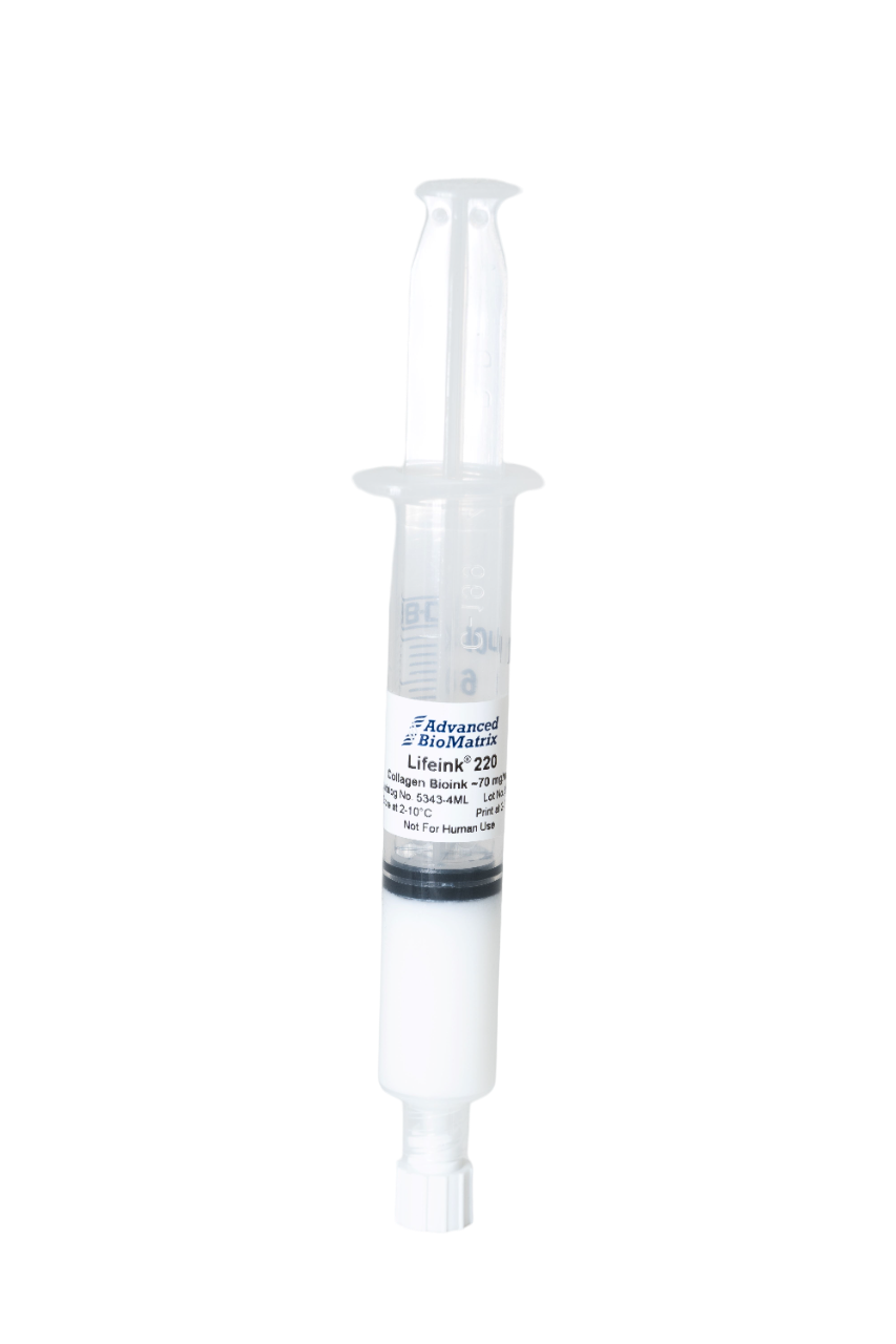
Lifeink® 220; Type I Collagen Bioink
Cat.-Nr: 5343-4ML
For printing with Lifeink® 220, we highly recommend using the FRESH printing technique and printing at 2-8°C. Three dimensional (3D) gels allow... Read More
-

Lifeink® 240, Acidic Type I Collagen Bioink
Cat.-Nr: 5267-5ML
Lifeink® 240 is an acidic Type I collagen bioink at a concentration of 35 mg/ml for extrusion-based 3D bioprinting. The product is a highly... Read More
-
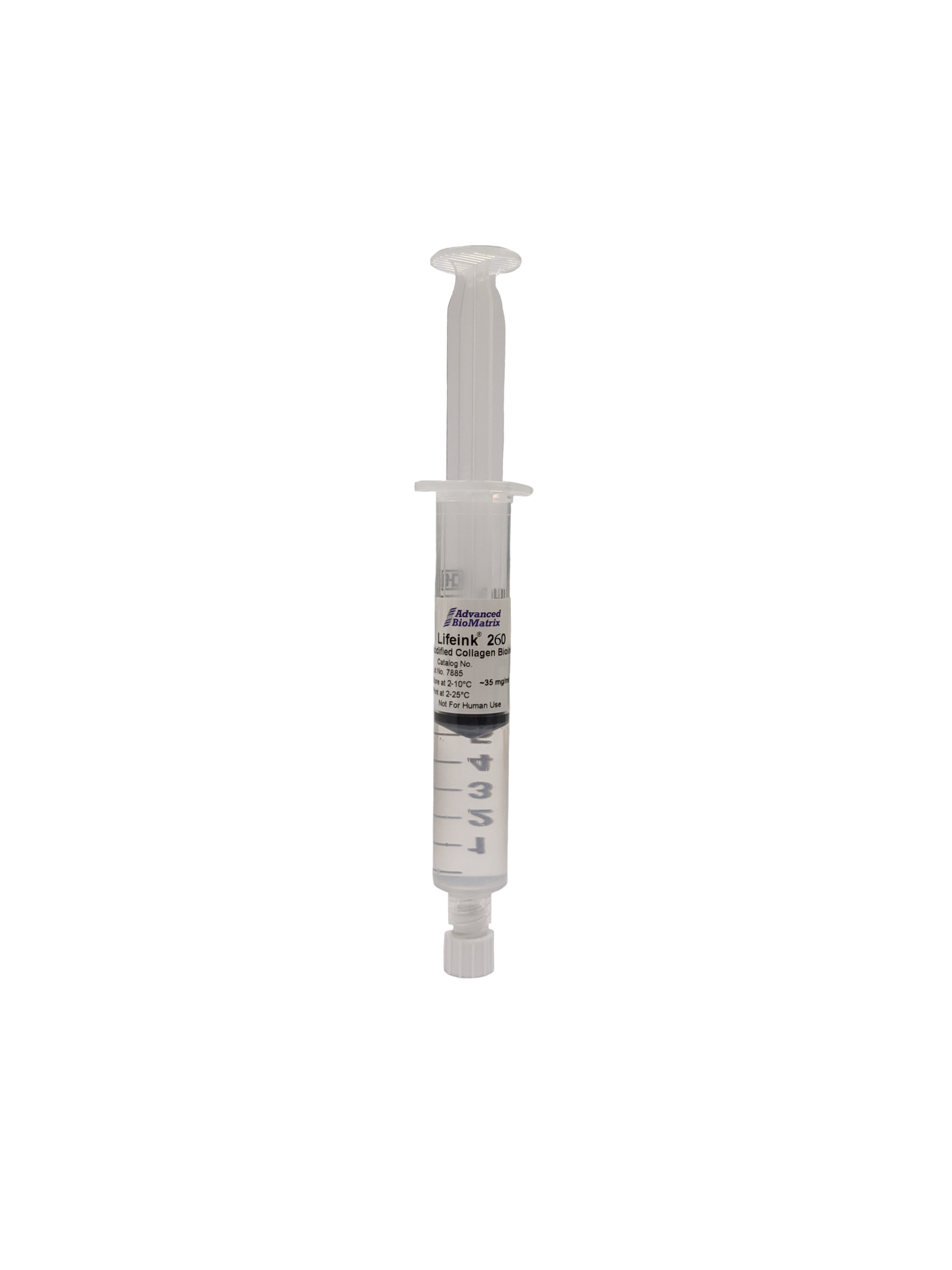
Lifeink® 260, Acidic Type I Collagen Bioink
Cat.-Nr: 5358-4ML
Lifeink® 260 is an acidic Type I collagen bioink at a concentration of 70 mg/ml for extrusion-based 3D bioprinting. The product is a highly... Read More
-
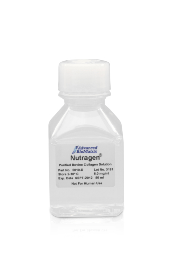
Nutragen®, Bovine Collagen, Solution
Cat.-Nr: 5010-50ML
Nutragen® collagen (bovine collagen, atelocollagen) is known as the standard of all collagens for purity (>99% collagen content), functionality,... Read More
-
Phosphate Buffered Saline
Cat.-Nr: 5076-100ML
Phosphate Buffered Saline (PBS) 10X is formulated and prepared to use in conjunction with other products provided by Advanced BioMatrix such as Type... Read More
-
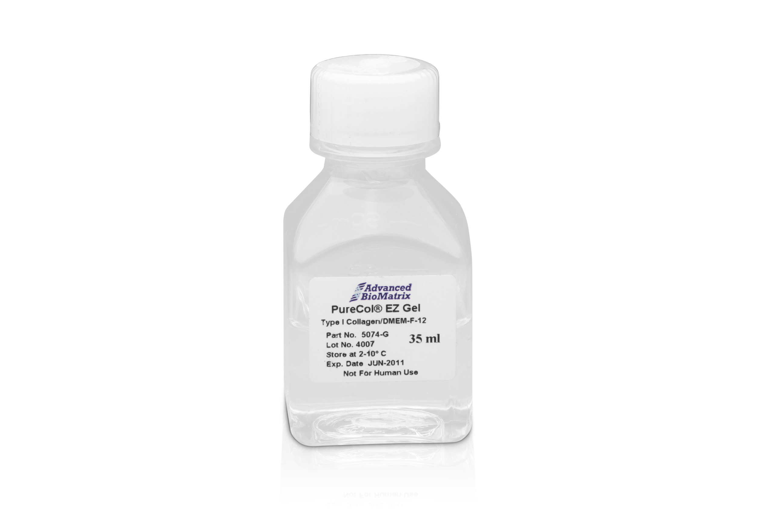
PureCol® EZ Gel, Bovine Collagen
Cat.-Nr: 5074-35ML
Important information: This product is currently not available for our customers in Europe. We offer as an alternative the Nutragen, bovine... Read More
-

PureCol®-S, Collagen Standard
Cat.-Nr: 5015-20ML
PureCol®-S collagen is provided as a standard for use in assays where ultra-pure collagen is required. PureCol®-S collagen standard is... Read More
-
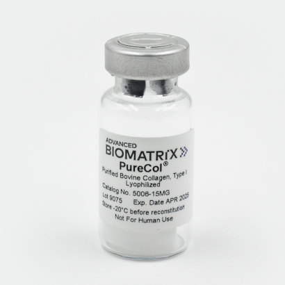
PureCol®, Bovine Collagen, lyoph.
Cat.-Nr: 5006-15MG
PureCol® collagen is known as the standard of all collagens for purity (>99.9% collagen content), functionality, and the most native-like... Read More
-
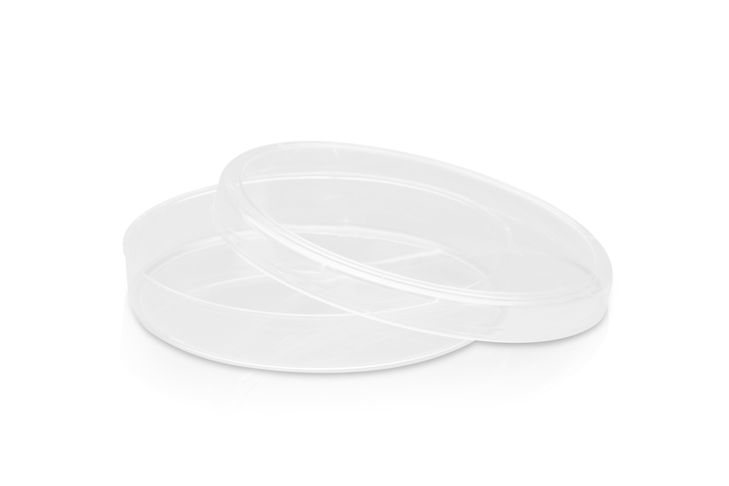
PureCol®, Collagen Coated, 100 x 20mm Dishes
Cat.-Nr: 5028-10EA
PureCol® Collagen Coated, 100 X 20 mm Dishes have a uniform and consistent application of high quality Type I collagen on clear polystyrene surface.... Read More
-
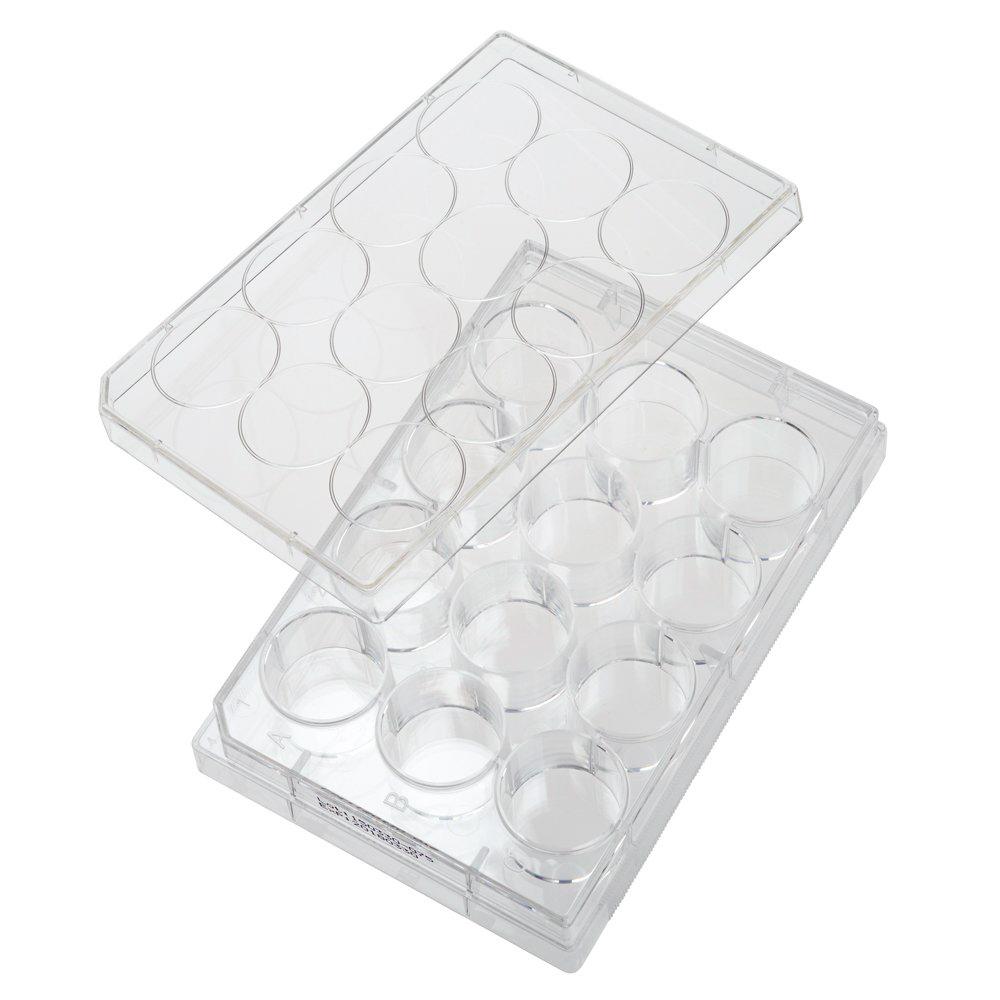
PureCol®, Collagen Coated, 12-well Plates
Cat.-Nr: 5439-5EA
PureCol® Collagen Coated, 12-well Plates with a flat bottom have an uniform and consistent application of high quality Type I collagen on clear... Read More
-
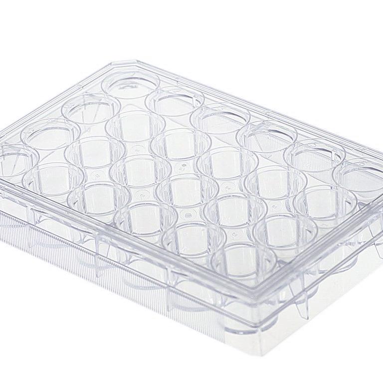
PureCol®, Collagen Coated, 24-well Plates
Cat.-Nr: 5440-5EA
PureCol® Collagen Coated, 24-well Plates with a flat bottom have an uniform and consistent application of high quality Type I collagen on clear... Read More
-
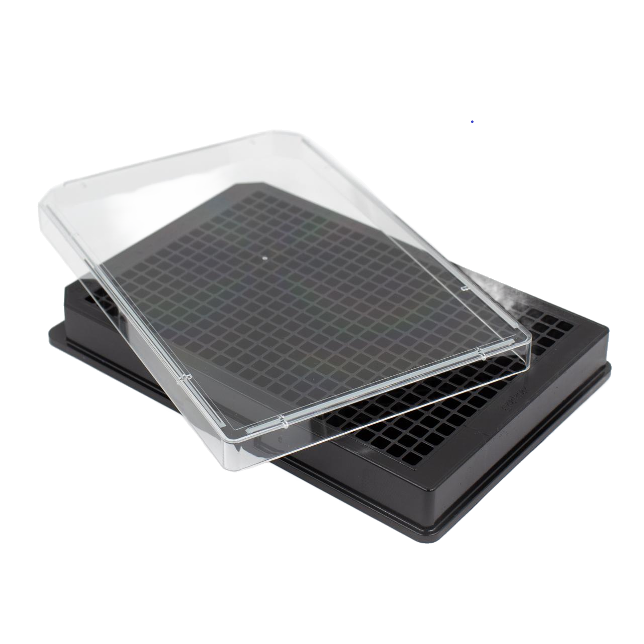
PureCol®, Collagen Coated, 384-well Plates
Cat.-Nr: 5380-5EA
PureCol® Collagen Coated, 384-well Plates have black walls and a flat bottom with a uniform and consistent application of high quality Type I... Read More
-
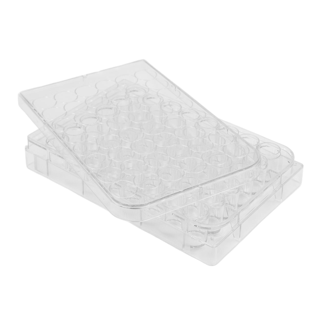
PureCol®, Collagen Coated, 48-well Plates
Cat.-Nr: 5181-5EA
PureCol® Collagen Coated, 48-well Plates with a flat bottom have an uniform and consistent application of high quality Type I collagen on clear... Read More
-
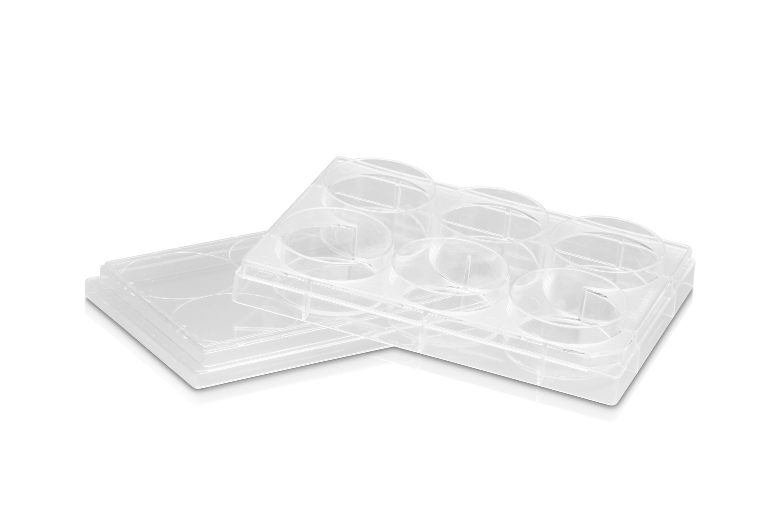
PureCol®, Collagen Coated, 6-well Plates
Cat.-Nr: 5073-5EA
PureCol® Collagen Coated, 6-well Plates with a flat bottom have a uniform and consistent application of high quality Type I collagen on clear... Read More
-
PureCol®, Collagen Coated, 96-well Plates
Cat.-Nr: 5072-5EA
PureCol® Collagen Coated, 96-well Plates with a flat bottom have an uniform and consistent application of high quality Type I collagen on clear... Read More
-
PureCol®, Collagen Coated, T25 Flasks
Cat.-Nr: 5029-10EA
Type III Collagen is typically used as a thin coating on tissue culture surfaces or as a control. Specific instructions are found in the Directions... Read More
-
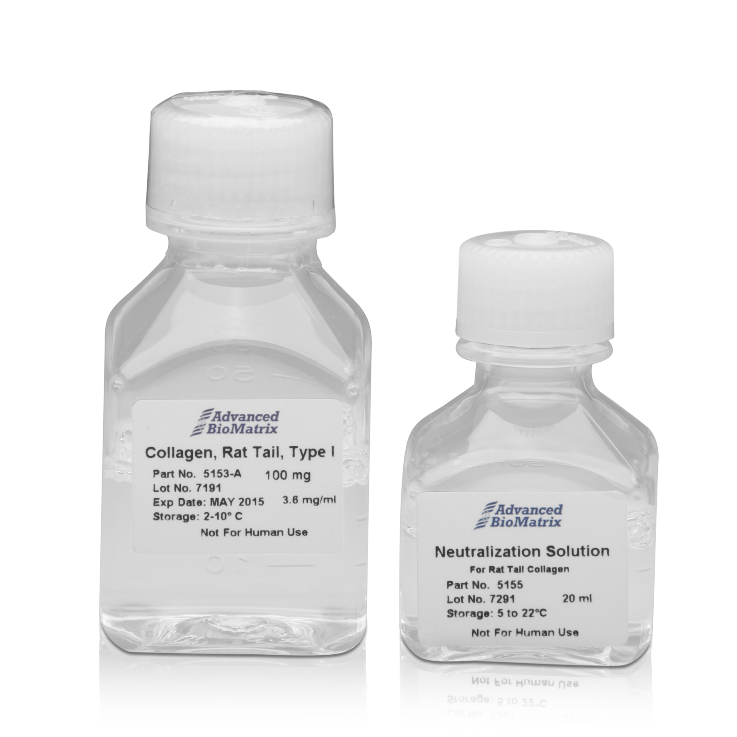
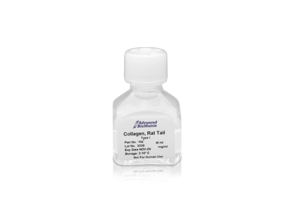
RatCol® Rat Tail Type I Collagen + Neutralizing Solution
Cat.-Nr: 5153-1KIT
RatCol® Type I Acid Soluble Rat Tail Collagen (telocollagen) contains 100 mg at a concentration of approximately 4 mg/mL in a 0.02M acetic acid... Read More
-
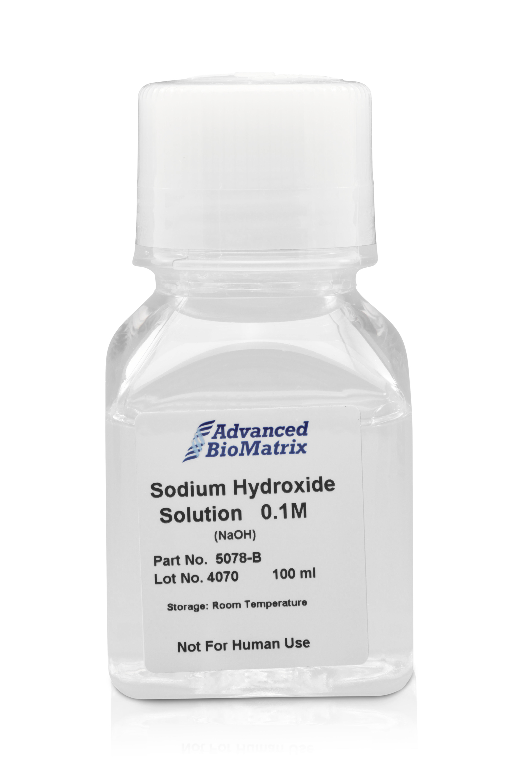
Sodium Hydroxide, Solution
Cat.-Nr: 5078-100ML
Sodium Hydroxide (NaOH) Solution 0.1M is formulated and prepared to use in conjunction with other products provided by Advanced BioMatrix. NaOH is... Read More
-
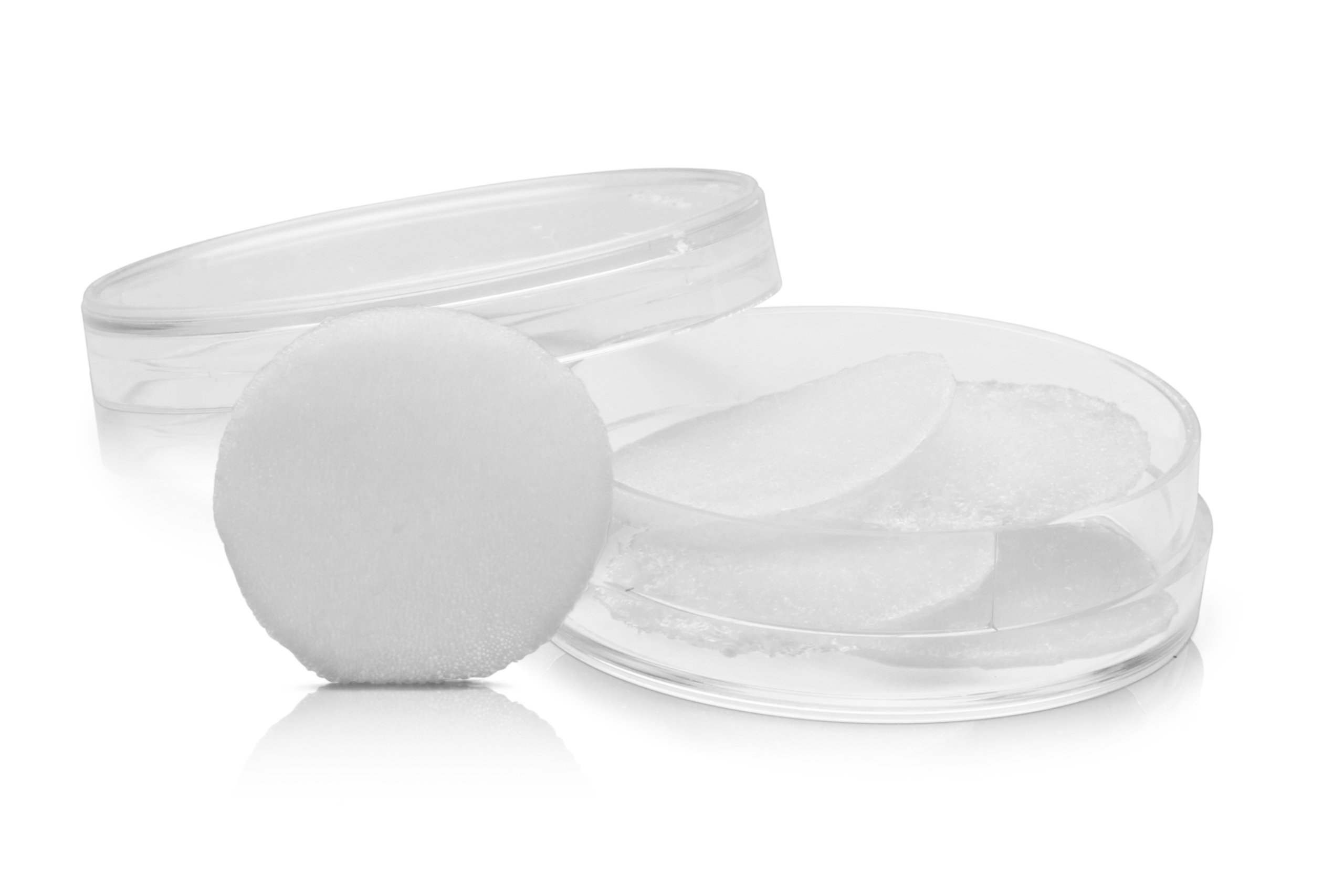
SpongeCol® Collagen Sponges with Columnar Pore Architecture, 21 mm Diameter Discs
Cat.-Nr: 5135-5EA
SpongeCol® is a collagen sponge with an interpenetrating, columnar porous architecture structure. SpongeCol® contains unique columnar porous... Read More
-
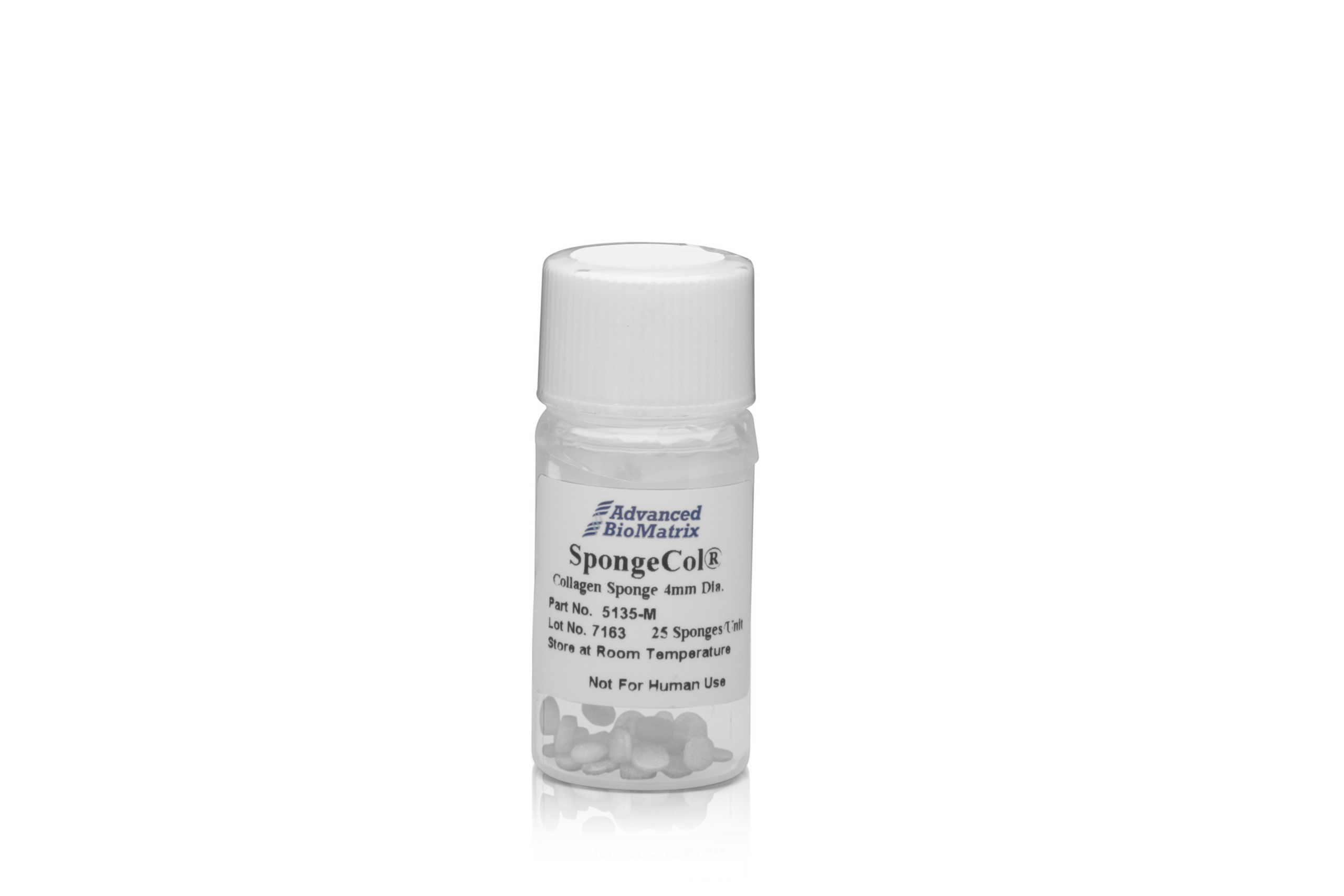
SpongeCol® Collagen Sponges with Columnar Pore Architecture, 4 mm Diameter Discs
Cat.-Nr: 5135-25EA
SpongeCol® is a collagen sponge with an interpenetrating, columnar porous architecture structure. SpongeCol® contains unique columnar porous... Read More
-

TeloCol®-10, purified Bovine Collagen, Solution
Cat.-Nr: 5226-20ML
TeloCol®-10 bovine collagen solution (telocollagen) is derived from an acid extraction process yielding a telopeptide-intact collagen. TeloCol® is... Read More
-

TeloCol™-3, purified Bovine Collagen, Solution
Cat.-Nr: 5026-1KIT
TeloCol®-3 bovine collagen solution (telocollagen) is derived from an acid extraction process yielding a telopeptide-intact collagen. TeloCol® is... Read More
-
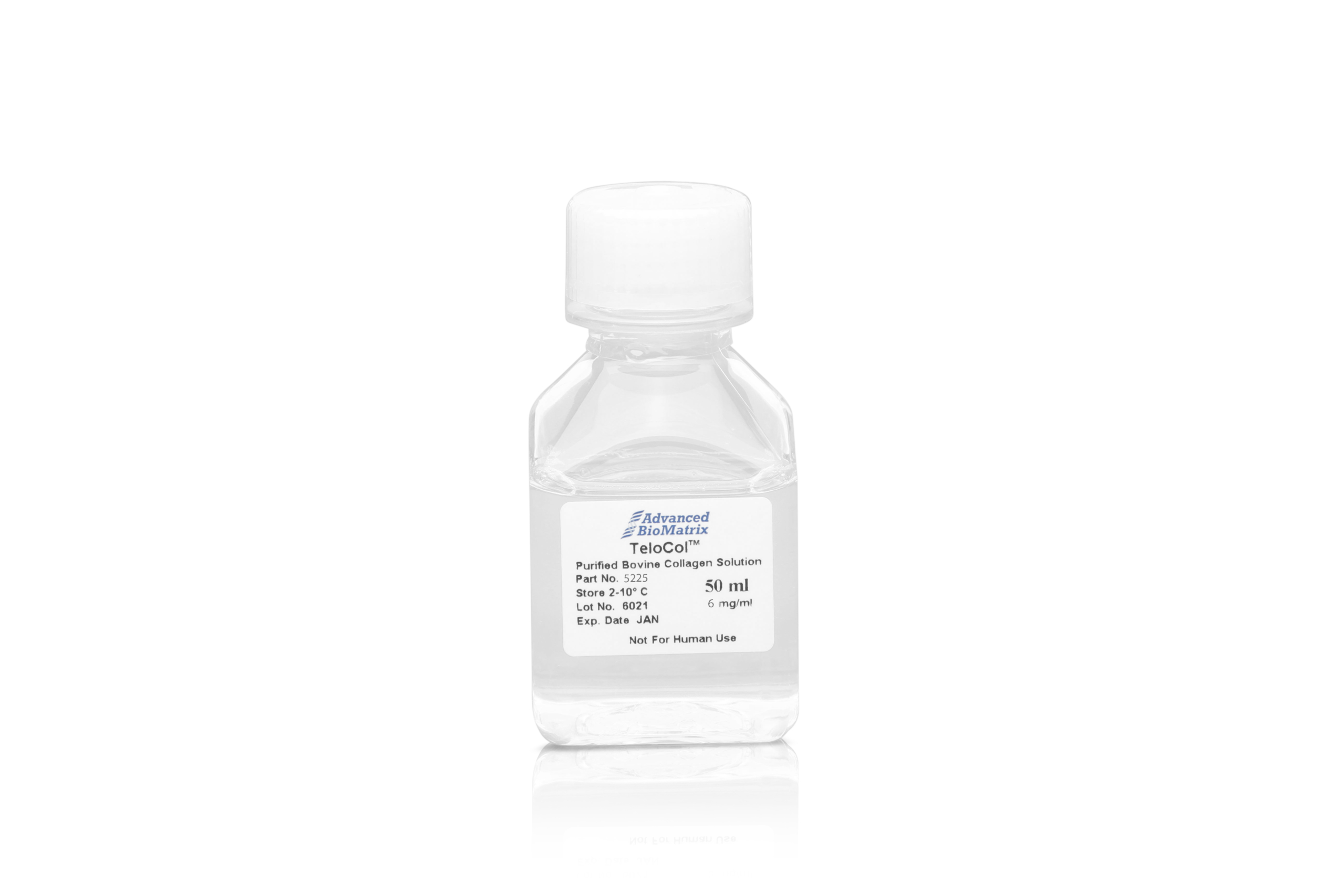
TeloCol™-6, purified Bovine Collagen, Solution
Cat.-Nr: 5225-1KIT
TeloCol®-6 bovine collagen solution (telocollagen) is derived from an acid extraction process yielding a telopeptide-intact collagen. TeloCol® is... Read More
-
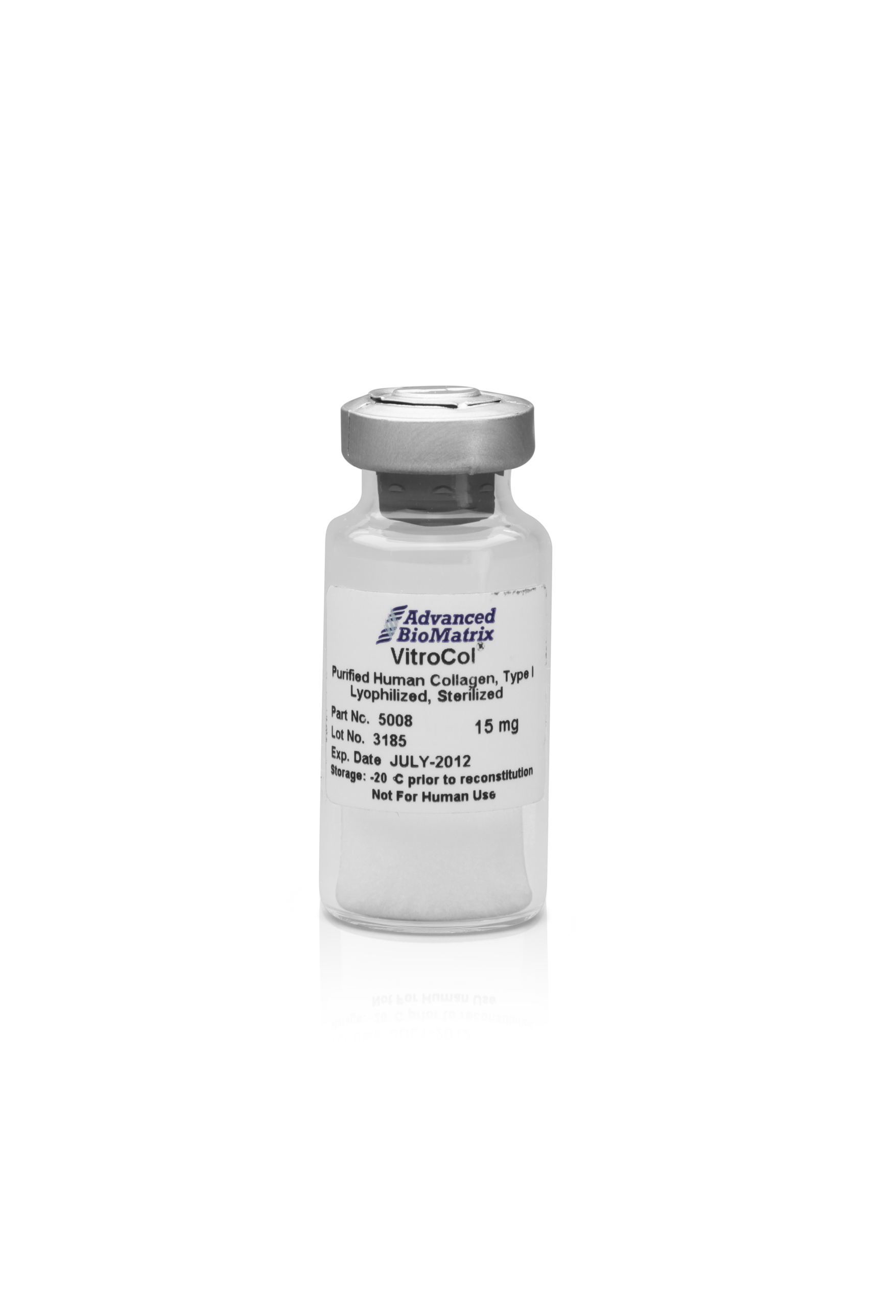
VitroCol®, Human Collagen, lyophilized
Cat.-Nr: 5008-15MG
VitroCol® collagen is the first widely available, naturally produced purified human collagen for research purposes. VitroCol® sets the standard for... Read More
-
VitroCol®, Human Collagen, Solution
Cat.-Nr: 5007-20ML
VitroCol® collagen is the first widely available, naturally produced purified human collagen for research purposes. VitroCol® sets the standard for... Read More