Next-generation full-thickness human skin models
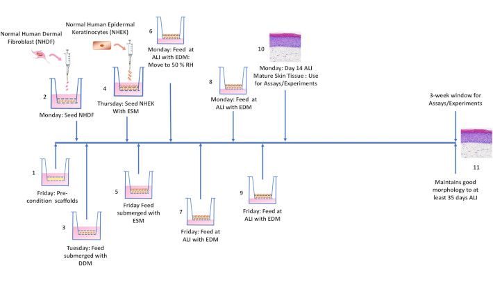
A newly published study in Frontiers in Toxicology demonstrates how electrospun 3D scaffolds can yield full-thickness human skin models that more closely recapitulate the structural and functional characteristics of native human skin than conventional hydrogel-based approaches – a meaningful step forward for toxicology testing, cosmetics research, and alternatives to animal testing. The models showed well-organized epidermal stratification, functional barrier properties, and dermal architecture comparable to ex vivo human skin.
The Scaffold: Biosurfaces Electrospun Matrices
Conventional full-thickness skin models typically rely on fibroblast-populated collagen or fibrin hydrogels as the dermal compartment. These matrix-based systems come with well-documented limitations: gel contraction driven by fibroblast-mediated remodeling compresses and distorts the dermal layer over time, shortens the usable lifespan of the model, and introduces batch-to-batch variability that makes reproducibility across experiments difficult to achieve.
The study directly addresses this by replacing the hydrogel with Bio-Spun® electrospun scaffolds from Biosurfaces . The authors show that fibroblasts cultured within the fibrous scaffold maintain a stable three-dimensional architecture without contraction, resulting in models with a significantly extended lifespan and consistent geometry across replicates. The electrospun fiber network — with diameters, porosity, and mechanical properties closely resembling native dermis — also supports more physiologically relevant fibroblast morphology and ECM deposition, and provides a structurally stable base for epidermal stratification. Barrier function and tissue architecture were validated against ex vivo human skin, with the models showing comparable performance across the evaluated parameters.
Biosurfaces electrospun scaffolds can be used directly as dermal substrates without modification or coating, making them a reproducible, off-the-shelf starting point for the dermal compartment.
Cells and Media: Building the Model with LifeLine®
The LifeLine portfolio combines primary human skin cells with tailored, phenol red-free culture media for reliable skin model generation. These physiologically relevant cell systems support applications ranging from tissue regeneration, pigmentation and cosmetic research to vascularization, drug permeation, and wound healing studies.
Build Your Next Skin Model with Confidence
Whether you are developing advanced 3D skin models for toxicology, cosmetic testing, wound healing, or dermatological research, the combination of Bio-Spun® electrospun scaffolds and well-characterized primary cell systems provide a robust foundation for reproducible full-thickness skin equivalents.
Contact us to discuss your application or request additional product information.
