2. Primary Cell Culture Problems: Causes & Solutions
Why Primary Cells Require Special Care
Primary cells closely mimic native tissue biology, making them invaluable for physiologically relevant research. However, their sensitivity to culture conditions means they require optimized handling, substrates, and media to achieve reliable attachment, survival, and proliferation. Success in primary cell culture depends on meeting cell-type-specific requirements and maintaining a stable microenvironment.
Poor growth and reduced viability in primary cell culture commonly result from:
-
Stress during isolation or thawing
-
Inadequate surface coatings that impair cell attachment
-
Suboptimal culture media lacking essential growth factors
-
Microbial contamination (bacterial, fungal, or mycoplasma)
Sterile Technique – Keeping Your Culture Clean and Reliable
Maintaining sterility in primary cell culture is non-negotiable. Even low levels of microbial contamination — whether bacterial, fungal, mycoplasma, or viral — can silently damage cell health, alter experimental outcomes, or force you to discard entire cultures.
Primary cells are especially vulnerable due to their limited lifespan and lack of selective growth advantages. Consistent aseptic technique, correct equipment use, and regular monitoring are essential to keep your cultures contamination-free.
Frequently Asked Questions
How can I maintain sterility and prevent contamination in primary cell culture?
-
Work inside a certified Class II biosafety cabinet (safety cabinet).
Turn on the safety cabinet at least 15–20 minutes before starting. Minimize movement and talking while working, as airflow disturbances can increase contamination risk. Disinfect all materials with 70% ethanol before placing them inside. -
Wear appropriate personal protective equipment: lab coat, gloves, and safety glasses if required.
Disinfect gloves regularly with 70% ethanol during the procedure to reduce microbial transfer.
- Prepare all necessary materials in advance:
- Prewarmed complete medium and PBS
- Sterile pipettes and tips
- Labeled flasks or dishes
- Waste container filled with disinfectant
-
Keep the work area organized.
Place only essential items inside the cabinet. Close bottles and tubes when not in use. Avoid placing caps or pipettes directly on surfaces. - Handle cells and reagents with sterile technique:
-
- Open flasks and pipette without touching sterile surfaces
- Aliquot reagents to prevent contamination of stock solutions
- Avoid passing hands or objects over open vessels
- When using trypsin, monitor detachment microscopically and neutralize promptly with serum-containing medium
-
Dispose of waste properly:
-
Collect liquid waste in a container with disinfectant inside the cabinet.
-
Discard solid waste into a sterile-lined biohazard container.
-
Clean all surfaces inside the cabinet with 70% ethanol after finishing.
-
-
After work:
Let the safety cabinet run for 10–15 minutes. Remove and disinfect all materials. Dispose of waste according to lab safety guidelines. Wash hands thoroughly.
Regularly monitor cell cultures for mycoplasma, bacterial, and fungal contamination. To help prevent bacterial and fungal contamination, the use of Gentamicin and Amphotericin B (GA) is recommended in eukaryotic cell cultures.
Many Lifeline® Complete Media Kits now include Gentamicin Amphotericin B as part of the included LifeFactors Kit. The inclusion of these antibiotics is optional and can be tailored to your specific experimental needs.
How frequently should I test my primary cell cultures for mycoplasma and other contaminants?
All Lifeline® normal and diseased human cells are confirmed negative for mycoplasma, bacterial, and fungal contamination upon delivery, providing a clean starting point for your cultures. However, contamination can still occur during routine handling. Therefore, it is important to test your primary cell cultures regularly – ideally upon initial thawing or isolation, and then at intervals of every 2 to 4 weeks during continued culture. Testing should also be performed before critical experiments or if you notice any unexpected changes in cell behavior or morphology. This routine testing helps maintain culture integrity and ensures reliable and reproducible results.
It is generally not recommended to culture cells routinely in antibiotics, as this can mask low-level contamination and potentially induce cell stress. Instead, maintain strict sterile technique and perform regular contamination testing to keep cultures clean without relying on antibiotics.
What are common sources of contamination and how can I avoid them?
Common sources of contamination in cell culture include:
-
Airborne microbes: Dust, spores, and bacteria from the environment can settle into open culture vessels.
-
Non-sterile equipment and reagents: Unsterilized pipettes, media, or reagents can introduce contaminants.
-
Poor aseptic technique: Touching sterile surfaces or cultures with ungloved hands or contaminated gloves.
-
Contaminated cell stocks or reagents: Using previously contaminated cells or reagents without proper testing.
-
Cross-contamination between cultures: Handling multiple cell lines without changing gloves or sterilizing tools.
-
Incubators and water baths: If not regularly cleaned and disinfected, they can harbor bacteria or fungi.
-
Personnel: Coughing, sneezing, or even a short chat and a little laughter while working at the bench passaging your cell cultures can introduce microbes, including mycoplasma.
When should I use antibiotics in my cultures, and what are the pros and cons?
Antibiotics can be used as a preventive measure to reduce bacterial contamination in cell cultures, especially during early stages or when working with primary cells that may be more vulnerable. They are also helpful when working in less-than-ideal sterile conditions or when contamination risks are high.
Pros:
-
Help prevent bacterial infections that could ruin cultures
-
Provide a safety net during routine handling or transport
-
Reduce the need for repeated culture discarding due to contamination
Cons:
-
May mask low-level contamination, making it harder to detect problems early
-
Can stress or alter cell physiology, affecting experimental outcomes
-
Risk of developing antibiotic-resistant contaminants
Many Lifeline® Complete Media Kits now include Gentamicin and Amphotericin B as part of the included LifeFactors Kit. The use of these antibiotics is optional and can be tailored to your specific experimental needs.
Whenever possible, it’s best to maintain strict aseptic technique and avoid routine antibiotic use. If you choose to use antibiotics, use them at the lowest effective concentration and for limited periods to minimize impact on your cells and experiments.
Thawing Primary Cells – Starting Off Right
Primary cells are much more sensitive than cell lines, and that sensitivity starts the moment you remove the vial from liquid nitrogen. You only get one chance to do this right.
One of the most critical steps in primary cell culture happens right at the beginning: thawing the cells. If the process is too slow, too rough, or poorly timed, even high-quality cells may fail to recover — and that can’t be undone.
Before thawing, make sure the cells have been stored correctly. Primary cells should always be kept in the vapor phase of liquid nitrogen. If you’re using vials that were stored in liquid phase, transfer them to the vapor phase or to –80 °C for no longer than 24 hours before thawing.
Frequently Asked Questions
What should I prepare before thawing primary cells?
Before you begin thawing, make sure the following steps are in place:
-
Wear proper protection: Always use gloves and eye protection when handling cryopreserved vials.
-
Prepare the culture medium as described in the datasheet: Follow all instructions carefully, including required supplements.
-
Pre-warm only what you need: If using less than 100 mL of medium, warm just the needed volume in a sterile conical tube. Avoid warming the entire bottle repeatedly — this shortens shelf life and degrades heat-sensitive components.
-
Don’t overheat: Medium will typically reach 37 °C in 10–30 minutes, depending on volume. Never leave it in the water bath for extended periods.
-
Vent the vial with the frozen primary cells just before thawing: Spray the vial with 70% ethanol or isopropanol and transfer it to a biosafety cabinet. Allow it to dry thoroughly and carefully loosen the cap to vent any liquid nitrogen that may have entered the vial. Then re-tighten until you’re ready to place the vial in the water bath.
With everything ready, you can now proceed with thawing — timing and handling are critical from here on.
What are the steps for thawing and plating primary cells?
- Recap the vial and hold only the bottom half in a 37 °C water bath for about one minute, or until the vial is almost completely thawed — a small amount of ice should still be visible.
Do not allow the vial cap to touch the water, to avoid contamination. Do not over-thaw, as this may damage the cells. - Dry the vial, spray the exterior with 70% ethanol or isopropanol, and place it in a biosafety cabinet to dry.
- Carefully remove the cap to avoid contamination or splatter.
- Gently resuspend the cells in the vial using a sterile 1- or 2-mL pipette.
- Take an aliquot (20 µL) to count viable cells using Trypan Blue and a hemacytometer (for the protocol, see the next question).
- Do not centrifuge the original vial; instead, plate the cells directly based on the viable cell number determined.
- Plate the cells at the recommended seeding density (e.g., 2.500 – 5.000 cells/cm²) into a T25 flask containing 5 mL of pre-warmed, fully supplemented medium specific to the cell type. Refer to the product data sheet for exact volumes and conditions. Use flasks with vented (filter) caps when possible.
- Gently rock the vessel side to side and front to back to distribute cells evenly.
- Place the vessel in a 37 °C, 5% CO₂ incubator.
- Refeed the cells after attachment (approximately 4 to 36 hours after seeding) to remove cryopreservation reagents.
How do I determine the cell number using Trypan Blue and a hemacytometer?
To perform a cell count using Trypan Blue and a hemacytometer:
- Resuspend your cells in the appropriate culture medium.
- Mix 20 µL of the cell suspension with Trypan Blue:
| Application | Recommended Dilution | Mix with 20 µL Cell Suspension | Notes |
|---|---|---|---|
| Initial Seeding | 1:2 | Add 20 µL Trypan Blue | Best for low cell densities where precise seeding is needed. |
| Routine Counting | 1:5 | Add 80 µL Trypan Blue | Works well for log-phase cultures; balanced visibility and accuracy. |
| High Cell Density | 1:10 | Add 180 µL Trypan Blue | Ideal when cultures are dense to avoid overloading the chamber. |
- Gently mix the Trypan Blue and cell suspension; avoid bubbles.
-
Let the mixture sit for 1–5 minutes to allow for accurate viability staining.
-
Count within 5 minutes of staining to minimize false positives.
-
Load 10 µL of the stained suspension into each chamber of a clean hemacytometer.
-
Count cells in 8 quadrants using standard inclusion rules:
-
Count cells touching the top or left border.
-
Do not count cells touching the bottom or right border.
-
-
Adjust cell concentration if too few (<120) or too many (>520) cells are counted.
-
Calculate viable cells per mL using the formula:
Viable cells/mL=(Total viable cells in 8 quadrants/8)×10,000*×dilution factor
*Note: The factor 10,000 converts the 0.1 µL volume of one quadrant to 1 mL.
How do I know if the cells are recovering properly?
-
Look for adherence, spreading, and typical morphology within the first 24–48 hours.
-
Avoid interpreting floating cells too early — some may still attach later.
-
Moderate cell death after thawing is normal; excessive floating debris may indicate DMSO shock, improper thawing, or plating issues.
-
Full confluence is not expected within the first days — focus on attachment and survival first.
When should I do the first medium change?
-
Wait until cells have clearly attached — typically 4 to 36 hours post-seeding, depending on the cell type.
-
A premature medium change may remove unattached but viable cells.
-
Watch for signs of attachment under the microscope (spread morphology, visible anchoring points).
-
Use gentle pipetting at the edge of the vessel when removing or adding medium.
- If unsure whether all cells have adhered, transfer the removed medium to a separate vessel and incubate for another 24 hours. Any viable, initially non-adherent cells can then attach, minimizing cell loss.
What are the basic steps for passaging adherent cells?
To passage adherent cells, follow these aseptic steps:
- Wash your hands and wear gloves and eye protection. Work in a certified biological safety cabinet.
- If serum-containing media is used, rinse each flask twice with Phosphate Buffered Saline (PBS) before adding 0.05% Trypsin/0.02% EDTA, such as that provided in TrypKit-100 ML (#LL-0013).
- Add the appropriate volume (see table below) of Trypsin/EDTA, and incubate for 1–3 minutes at room temp or 37°C, depending on cell type. Observe with a microscope.
Note: Some strongly adherent cell types, such as keratinocytes, may require double the recommended volume, significantly longer incubation time, or trypsinization at 37 °C.
Note: Over-trypsinaization may damage cells.
| Product | Volume | Storage | Volumes to Use | ||
|---|---|---|---|---|---|
| per 1 cm2 | per T25 flask | per T75 flask | |||
| TrypKit (#LL-0013) | Kit | ||||
| PBS without calcium or magnesium | 500 mL | RT | 0.2 mL | 5 mL | 15 mL |
| 0.05% Trypsin/0.02% EDTA | 100 mL | -20°C | 0.02 mL | 0.5 mL | 1.5 mL |
| Trypsin Neutralizing Solution (TNS) | 100 mL | -20°C | 0.02 mL | 0.5 mL | 1.5 mL |
Gently tap the flask to detach cells once they begin lifting. Some cell types may require more tapping or longer incubation.
- Add an equal volume of TNS and mix to neutralize the trypsin/EDTA, and transfer cells to a sterile centrifuge tube.
- Rinse the culture vessel with PBS to collect any remaining cells, and combine in the same tube.
- Check culture vessel under the microscope for cells still attached and repeat steps if necessary to retrieve all the cells from the vessel.
- Centrifuge cells at 150 x g* for 3–5 minutes. Aspirate supernatant carefully.
- Re-suspend the cell pellet gently in pre-warmed (37°C) culture medium.
- Add medium to new flasks (e.g., 1 mL per 25 cm²) and seed cells according to your Cell Instruction Sheet.
Always avoid over-trypsinizing or over-centrifuging, and calculate RCF appropriately for your centrifuge model.
*To calculate RCF (x g): RCF = 0.00001118 × (rpm)2 × r
r = rotational radius in centimeters
rpm = rotations or revolutions per minute
Poor Cell Attachment – Common Causes and Fixes
Primary cells often struggle to adhere to standard tissue culture plastic, which can result in reduced viability, slow proliferation, or early cell loss. To promote proper attachment and support cell-specific behavior, it is often necessary to coat the culture surface with extracellular matrix (ECM) proteins or synthetic substrates. This step is particularly important when working with serum-free or defined media, which lack naturally occurring adhesion factors.
ECM coatings help recreate the cells’ natural microenvironment, providing critical signals for adhesion, survival, and function. The optimal coating should reflect the native niche of the respective cell type—for example, fibronectin supports endothelial cells, while neurons require poly-D-lysine and laminin.
The following section offers practical guidance on selecting appropriate coatings for a variety of primary cell types.
| Primary Cell Type | Preferred Coating / Attachment Factors | Rationale & Notes |
|---|---|---|
| Fibroblasts (e.g., dermal, neonatal) | Collagen Type I, Fibronectin | Fibroblasts naturally interact with collagen-rich matrices and adhere efficiently to ECM-coated surfaces. |
| Endothelial Cells (e.g., HUVEC, HMVEC) | Fibronectin, Collagen Type I, Vitronectin | Supports monolayer formation, barrier function, and cell–cell interactions. Vitronectin can enhance adhesion in serum-free systems. |
| Mesenchymal Stem Cells (MSC) | Collagen Type I, Fibronectin, Vitronectin | Support adhesion and influence lineage-specific differentiation. |
| Hepatocytes (primary, human/rat) | Collagen Type I | Helps maintain polarity and functional hepatocyte morphology in vivo. Sandwich cultures (collagen overlays) further support functionality. |
| Neurons (e.g., cortical, DRG) | Poly-D-lysine + Laminin or Collagen Type I | Essential for axonal growth and maturation; fibronectin supports progenitor adhesion. |
| Glial Cells (astrocytes, microglia) | Fibronectin, Laminin, Collagen Type I | Promote adhesion and preserves glial morphology and function. |
| Keratinocytes (primary, human) | Collagen Type IV, Fibronectin, Vitronectin | Mimics basement membrane, critical for proper differentiation and attachment. |
| Myoblasts / Skeletal Muscle Progenitors | Collagen Type I, Laminin, Fibronectin | Collagen supports alignment and fusion during myogenesis. |
| Chondrocytes | Collagen Type II (or Type I), Fibronectin | ECM-mimicking coatings help maintain chondrogenic phenotype and adhesion. |
| Pulmonary Epithelial Cells (e.g., SAEC) | Collagen Type I, Fibronectin | Support differentiation and tight junction formation in air liquid interface (ALI) cultures. |
| iPSCs / ESCs (differentiating) | Vitronectin, Laminin, Tropoelastin | Defined substrates that maintain pluripotency and enable reproducible differentiation protocols. |
Frequently Asked Questions
How do I coat plates for improved cell attachment?
Common coatings used in cell culture include collagen, various extracellular matrix proteins such as fibronectin, and adhesion-promoting peptides like poly-D-lysine. The optimal choice depends on the cell type and application.
Detailed instructions and directions for use with coating protocols are available for download on each product page.
Coating Procedere
Note: Use these recommendations as guidelines to determine the optimal coating conditions for your culture system.
Note: Employ aseptic practices to maintain the sterility of the product throughout the preparation and handling of the extracellular matrix protein or attachement factor and other solutions. To maintain sterility, perform all operations in a laminar flow hood.
Note: Allow all materials and vessels to equilibrate to room temperature before use.
- Prepare working solution:
Transfer the required volume of ECM or adhesion reagent into a sterile dilution vessel. Dilute to the desired working concentration using the appropriate diluent (see table below).
| Product | Typical Working Concentration | Diluent | Rinsing Solution |
|---|---|---|---|
| PureCol® (Bovine Collagen Type I) | ~50–100 µg/mL (~1:30) | Cell culture grade water or 0.01 N HCl | Sterile Medium or PBS |
| RatCol™ (Rat Tail Collagen Type I) | ~50–100 µg/mL | 0.1% Acetic Acid | Sterile Medium or PBS |
| Human Collagen Type III | 50–100 µg/mL (~1:10) | 0.01 M HCl in cell culture grade water | Sterile Medium or PBS |
| Fibronectin (Human) | PBS or serum-free medium (Ca²⁺/Mg²⁺-free) | Sterile distilled water | |
| Poly-D-Lysine (PDL) | ~100 µg/mL | Cell culture grade water or PBS | Cell culture grade water |
- Apply to culture surface:
Add enough volume to completely cover the surface of the culture vessel. Example: 1 mL per 25 cm² (T25 flask or equivalent well area). Gently tilt to ensure even coating. - Incubate:
Incubate the coated vessel at room temperature for 1–2 hours (or until dry, if applicable). Cover to prevent contamination.
Note: the incubation time for PDL is only 5 minutes. - Remove excess coating solution:
Aspirate remaining liquid without scratching the surface. - Rinse (if required):
Rinse gently with sterile rinsing solution (see table above). Do not disturb the coated layer. - Storage or immediate use:
Use immediately or store coated vessels at 2–8 °C (damp or air-dried) if sterility is maintained. Coated cultureware can be stored for up to 1 week (PDL) or longer. To air-dry, incubate at room temperature or 37°C, covered, for 1-2 hours.
How can I identify the best extracellular matrix coating for my cells?
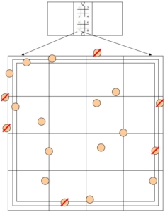
To help determine the most effective coating for your specific cell type, we recommend using the ECM Select® Array Kit Ultra-36, which enables side-by-side comparison of 36 different extracellular matrix and adhesion protein combinations.
What are the key differences between 2D and 3D cell culture?
-
2D Culture: Cells are grown as a monolayer on a flat surface, which is easier to handle and analyze but may not represent native tissue architecture.
-
3D Culture: Cells are grown within or on top of a matrix, forming spheroids or organoids, better mimicking in vivo conditions and cell interactions but are technically more challenging.
When should I use phenol red in cell culture media, and why might I choose not to?
Phenol red is a pH indicator added to culture media to help visually monitor pH changes—it turns yellow in acidic and purple in basic conditions. However, it is not required for cell culture and may adversely affect the behavior of some sensitive cell types or interfere with certain assays. If you want to add phenol red to Lifeline® media, you can purchase it separately (LS-1009) and add 0.5 mL per 500 mL of medium to achieve a typical concentration of 33 µM. The supplement is stable at room temperature. Use phenol red only if pH monitoring by color change is important and if it does not impact your cells or experiments.
Poor Cell Proliferation – Causes and Corrective Actions
Even when cells attach and survive the thawing process, inappropriate medium composition can silently impair growth, function, or morphology. Primary cells rely on precisely balanced nutrients, growth factors, and pH — and unlike immortalized lines, they rarely tolerate deviation. This section helps you identify signs of media-related issues and how to correct them.
What are signs that my medium might not be suitable?
-
Cells grow slowly or stop proliferating altogether.
-
Morphology looks abnormal (e.g., overly rounded, vacuolated, or flattened).
-
Cell detachment or spontaneous death increases over time.
-
Differentiation markers are lost or altered.
Frequently Asked Questions
Why are my primary cells not growing well in culture?
Primary cells are more sensitive than immortalized cell lines and require optimized conditions for attachment, survival, and proliferation. Poor growth may result from:
suboptimal medium formulation,
lack of specific growth factors,
inappropriate surface coating,
stress during isolation or thawing, or
contamination.
Ensure that culture conditions match the cell type’s physiological needs and that handling is gentle and aseptic throughout.
How does the choice of culture medium affect proliferation rates?
The choice of culture medium is critical for cell proliferation because it provides essential nutrients, growth factors, and signaling molecules that support cell survival and division. Using a medium that is specifically formulated for your primary cell type ensures optimal conditions for growth. Suboptimal or generic media may lack key components, leading to slower proliferation or poor viability. With Lifeline® media, you’ll get consistently reproducible results—each medium is carefully balanced and supplemented expressly for a particular primary cell type to maximize that cell type’s growth and longevity.
Could my cells be nearing senescence, and how can I tell?
Primary cells have a limited lifespan and eventually enter senescence, a state where they stop dividing and show altered morphology and function. Signs of senescence include slowed proliferation, enlarged and flattened cell shapes, increased granularity, and changes in gene expression. Monitoring population doublings and passage number is important, as cells closer to their limit will show these features. Using cells within the recommended population doubling range and at early passages can help you avoid working with senescent cultures.
How can population doublings be used to track cell proliferation capacity?
Population doublings (PDs) measure how many times a cell population has doubled during culture and provide a precise way to track cell proliferation capacity over time. By calculating PDs at each passage, you can monitor the growth potential and detect when cells begin to slow down or approach senescence. Keeping track of PDs helps ensure you use cells within their optimal proliferative lifespan for reliable and reproducible results.
To calculate population doublings (PD), use this formula:
PD=(log(N f)−log(N i))log(2)
where:
- NiN i = number of cells you initially plated
- NfN f = number of cells you harvested at the end of the culture period
This calculation tells you how many times the cell population has doubled between plating and harvest. Keep track of PDs cumulatively across passages to monitor overall cell proliferation capacity.
Routine Maintenance of Primary Cell Cultures
Before regular feeding and passaging routines begin, freshly thawed adherent cells must first attach firmly to the culture vessel surface. Successful attachment within the first 4–36 hours is critical for survival and uniform growth. Once cells are attached and have resumed normal morphology, the focus shifts to consistent care – maintaining optimal density, nutrient levels, and environmental conditions.
Routine culture work is not just about feeding and splitting cells; it’s about preventing stress, contamination, and unwanted variation to keep cells healthy and experimental results reproducible.
Frequently Asked Questions
What are the basic steps for passaging adherent cells?
To passage adherent cells, follow these aseptic steps:
-
Wash your hands and wear gloves and eye protection. Work in a certified biological safety cabinet.
- If serum-containing media is used, rinse each flask twice with Phosphate Buffered Saline (PBS) before adding 0.05% Trypsin/0.02% EDTA, such as that provided in TrypKit-100 ML (#LL-0013).
-
Add the appropriate volume (see table below) of Trypsin/EDTA, and incubate for 1–3 minutes at room temp or 37°C, depending on cell type. Observe with a microscope.
Note: Some strongly adherent cell types, such as keratinocytes, may require double the recommended volume, significantly longer incubation time, or trypsinization at 37 °C.
Note: Over-trypsinaization may damage cells.
| Product |
Volume |
Storage |
Volumes to Use |
||
| per 1 cm2 | per T25 flask |
per T75 flask |
|||
| TrypKit (#LL-0013) |
Kit |
||||
|
PBS without calcium or magnesium |
500 mL | RT | 0.2 mL | 5 mL |
15 mL |
|
0.05% Trypsin/0.02% EDTA |
100 mL | -20°C | 0.02 mL | 0.5 mL |
1.5 mL |
| Trypsin Neutralizing Solution (TNS) |
100 mL |
-20°C | 0.02 mL | 0.5 mL |
1.5 mL |
Gently tap the flask to detach cells once they begin lifting. Some cell types may require more tapping or longer incubation.
-
Add an equal volume of TNS and mix to neutralize the trypsin/EDTA, and transfer cells to a sterile centrifuge tube.
-
Rinse the culture vessel with PBS to collect any remaining cells, and combine in the same tube.
- Check culture vessel under the microscope for cells still attached and repeat steps if necessary to retrieve
all the cells from the vessel. -
Centrifuge cells at 150 x g* for 3–5 minutes. Aspirate supernatant carefully.
-
Re-suspend the cell pellet gently in pre-warmed (37°C) culture medium.
-
Add medium to new flasks (e.g., 1 mL per 25 cm²) and seed cells according to your Cell Instruction Sheet.
Always avoid over-trypsinizing or over-centrifuging, and calculate RCF appropriately for your centrifuge model.
*To calculate RCF (x g): RCF = 0.00001118 × (rpm)2 × r
r = rotational radius in centimeters
rpm = rotations or revolutions per minute
What is the recommended seeding density for primary adherent cells?
Optimal seeding density depends on the cell type but generally ranges from 2,500 to 5,000 cells/cm². Primary cells should not be seeded too sparsely, as they often rely on cell–cell signaling for proper attachment and proliferation. Please refer to the datasheet specific to your cell type.
Human mesenchymal stem cells (HMSCs), for example, should be seeded at higher densities—typically between 5,000 and 20,000 cells/cm².
How often should I change the medium in primary cell culture?
Medium should generally be changed every 2 to 3 days, depending on cell density and metabolic activity. As the medium does not contain a pH indicator, monitor culture health by observing cell morphology and confluence under the microscope. If visual pH monitoring is desired, Phenol Red (#LS-1009) can be added separately at 0.5 mL per 500 mL of medium (final concentration: 33 µM).
How do I know when my cells are ready to be passaged?
Most normal human adherent cells should be passaged when they reach 70–100% confluence and are still actively proliferating (evidenced by the presence of mitotic figures). For guidance on the appropriate density and confluence for your cell type, refer to the cell-specific Lifeline® Cell Technologies Instruction Sheets, which typically include reference images on the front page. Depending on the cell type, recommended split ratios generally range from 1:3 to 1:12.
How long can I culture human primary cells?
With Lifeline® media, primary cells can generally be maintained for at least 15 population doublings. Note that population doublings measure the actual number of times the cells have doubled, which differs from the number of passages or splits. This distinction is important for tracking cell aging and experimental consistency.
What is the difference between passage number and population doublings in primary cell culture?
Passage number tracks how often cells are subcultured, but it doesn’t reflect actual cell proliferation. In contrast, population doublings (PDs) quantify how many times the cell population has doubled and are a better measure of the culture’s biological age — especially important for primary cells, which have a limited replicative lifespan.
One passage typically corresponds to approximately 1–3 population doublings, depending on seeding density, growth conditions, and cell type. For primary cells, this means that the theoretical replicative capacity (often around 15 doublings for many human primary cultures) can be substantially consumed within the first few passages. As a result, primary cultures generally reach their biological limits by passage 3–4, after which further expansion often leads to reduced proliferative capacity, morphological changes, and altered functionality. Passages beyond P5 are therefore uncommon for primary cells and may indicate culture adaptation or selection effects.
To calculate PDs:
For example, if you seed 1 × 10⁵ cells and harvest 8 × 10⁵ cells:
Since PDs accumulate over time, tracking cumulative PDs gives a more accurate picture than passage number alone.
In contrast, immortalized or tumor-derived cell lines can tolerate much higher passage numbers (e.g., P10–P20 and beyond) with relatively stable proliferation, although genetic drift and phenotypic changes may still occur over extended culture.
When should I use phenol red in cell culture media, and why might I choose not to?
Phenol red is a pH indicator commonly used in cell culture media to provide a visual cue of pH changes—appearing yellow under acidic and purple under basic conditions. While useful for monitoring, it is not essential and may interfere with sensitive cell types or specific assays.
Use Phenol Red (#LS-1009) only if visual pH monitoring is important and its presence does not impact your cells or experimental outcomes. Add 0.5 mL per 500 mL of medium to achieve a final concentration of 33 µM.
Why can purity decrease during culture?
Although starting cultures are highly enriched for the target cell type, minor populations of accessory or co-isolated cells may be present. Over time, these cells can proliferate under culture conditions and alter the population balance — a known consequence of the biological heterogeneity of primary cultures and subpopulation dynamics during long-term maintenance.
Cryopreservation and Extended Culture Management
Cryopreservation is an essential component of extended culture management, enabling long-term storage of primary cells while preserving valuable biological material for future use. Although frozen cells can be recovered for subsequent experiments, cryopreservation affects the remaining proliferative capacity of primary cultures and requires optimized protocols to ensure viability and functional recovery. The following sections address key considerations, frequently asked questions, and best practices for successful cryostorage and post-thaw handling.
Frequently Asked Questions
Can I freeze my cultured primary cells?
Yes, cultured primary cells can be cryopreserved for long-term storage and experimental flexibility using solutions optimized for the cryopreservation of primary mammalian cells, such as FrostaLife™ Cryopreservation Solution, a ready-to-use freezing solution. After counting and centrifuging the cells, simply resuspend the pellet in FrostaLife™ Cryopreservation Solution and transfer to cryovials for storage. FrostaLife contains DMSO and fetal bovine serum, but no antimicrobials or phenol red. Each production lot is manufactured under ISO-style quality standards and tested for performance using primary human melanocytes, ensuring consistent and reproducible results.
Do I lose doubling capacity when freezing primary cells?
Yes. Primary cells have a finite number of population doublings. For example, primary human cells supplied by Lifeline Cell Technology are typically characterized by approximately 15 total doublings. Cryopreservation does not reset this limit; it consumes part of the available capacity — often reducing remaining doublings by roughly 2–3 in addition to those already used before freezing.
Are all primary cell types suitable for cryopreservation?
No. Cryotolerance varies by cell type:
-
Well suited: fibroblasts, keratinocytes
-
Less suited: specialized or fragile cell types (e.g., fallopian epithelial cells and similar lineage-specific cultures)
Less cryotolerant cells may show reduced post-thaw viability or functional performance. In such cases, continuous culture or alternative strategies may be preferable.
Which cryopreservation medium should I use?
Defined cryopreservation solutions optimized for primary mammalian cells are recommended, such as FrostaLife™ Cryopreservation Solution. These media typically contain DMSO as a cryoprotectant and may include serum components but do not contain antimicrobials or phenol red.
Do frozen primary cells retain biological function after thawing?
In most cases, yes — provided that cryopreservation and thawing protocols are properly executed. However, some functional or viability differences may occur, particularly with sensitive cell types. Careful optimization of freezing and recovery conditions improves reproducibility and performance.