We are here to help   +49 2241 25515 0
  • Shopping Cart Shopping Cart
    0Shopping Cart
  • Products
    • Primary Cells & Culture Media
      • Primary Cells
      • Growth Media
      • Differentiation Media
      • Subculture & Reagents
    • Tissue Dissociation
      • Collagenase & Dispase
      • Papain & Trypsin
      • Elastase & Hyaluronidase
      • Tissue Dissociation Kits
    • Matrix Proteins / Bioprinting
      • Collagen
      • Extracellular Matrix Proteins
      • Tunable Stiffness
      • Adhesion Molecules & Peptides
    • iPSC Research
      • iPSC-Derived Cells
      • MEFs
      • Media & Reagents
      • Services & Products
      • iPSC Lines
    • Other Products
      • Enzymes
      • Natural Proteins
      • Staining Kits
      • Cultureware
    • Cell Signaling
      • Cytokines
      • Chemokines
      • Growth Factors
      • Neurotrophins
      • CD Antigens
    • Molecular Biology
      • DNase & Related Products
      • RNase & Related Products
      • Enzymes & Related Products
    • Antibodies
      • Coagulation
      • Immunology
      • Cell Biology
    • Haemostasis & Thrombosis
      • Coagulation Proteins
      • Factor Deficient Plasmas
      • Bone Related Proteins
      • Fluorogenic Substrates & Inhibitors
      • Sample Collection Tubes
  • Services
  • Brands
  • Company
  • Career
  • News
  • Contact
  • Downloads
    • Portfolio Flyer
  • Menu Menu
Home |Products|Matrix Proteins / Bioprinting|Tunable Stiffness|Silk Fibroin Solution, 50 mg/ml

Silk Fibroin Solution, 50 mg/ml

Cat.-Nr.: 5154-20ML

Description

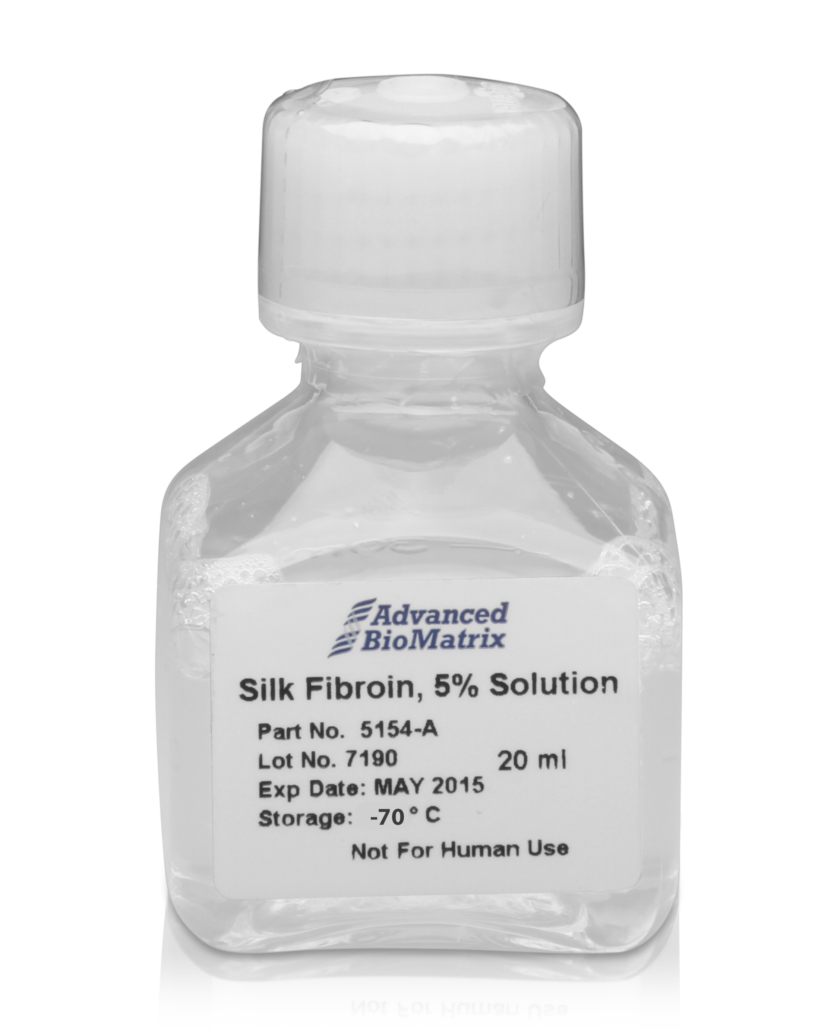
Advanced BioMatrix’s silk solution is approximately 50 mg/mL (5% W/V) of solubilized protein with a molecular weight of approximately 100k Da, available in 20 mL volume. The silk solution is made of 100% fibroin protein that is derived from the domesticated Bombyx mori silkworm. The product is manufactured in a manner to minimize contamination and has a low bioburden but is not considered sterile.

Fibroin protein is the major structural component of the silkworm’s cocoon fiber. Fibroin offers great potential for use in medically related applications due to the high degree of biocompatibility and lack of immune response when implanted within the body. The silk fiber is solubilized into an aqueous fibroin solution, which can then be used as an additive in culture or for producing 3D scaffolds for tissue-engineering related studies.

As with traditional tissue-engineering approaches, the silk scaffolds are typically seeded in vitro with a specific cell type as most cells will adhere to fibroin protein, and then cultured over time to mimic tissue architecture. It has been shown that the silk fibroin protein can be degraded a number of naturally occurring proteolytic enzymes, and is thus a biologically active scaffold unlike other synthetic materials. As a result the silk scaffold material is degraded and remodeled through similar physiological pathways in the body. Silk fibroin protein is composed of both non-essential and essential amino acids, with a particular concentration of alanine and glycine present, and these amino acids are then reabsorbed by the surrounding cells for new tissue regeneration. This is important as silk degradation products do not collect in the local environment to induce a toxicity which is commonly associated with other synthetic and naturally occurring biomaterials.

The ability to produce a variety of forms and formats scaffold types (e.g. coatings, films, sponges, hydrogels, electro-spun fibers, micro/nanospheres, etc.) offers a number of advantages over other biopolymer systems like collagen, chitosan, and alginate that have less variety in processing choices. The silk material properties can then be modified through a variety of processing techniques to change degradation rate, hydrophobicity/hydrophilicity, transparency, mechanical strength, porosity, oxygen permeability, and thermal stability. In this regard, silk proteins represent a class of biopolymers with definable material properties for a given application.

This product is prepared from silk fibroin extracted from Bombyx mori silkworm cocoons and contains a high monomer content with a molecular weight of approximately 100k Da. It is supplied as a ~50 mg/mL (5%) aqueous solution. This product is aseptically processed resulting in a low bioburden but is not considered sterile. If culturing cells using this product, measures should be taken to maintain sterility of cultures such as use of antibiotics.


The prodcut is available as a lyophilized powder, as well – (CytoSilk Silk Fibroin – CellSystems®)


  • SUPPLIER:

    Advanced BioMatrix

  • STATUS:

    In Stock

  • SIZE:

    20 ml

  • Overview
  • Related Files
  • References

Overview

  • Species: Other
  • Keywords: 3D Hydrogels, Reagents
  • Features:50 mg/ml

Related Files

Datasheet
Certificate of Origin

References

  • Compaan, Ashley M., Kyle Christensen, and Yong Huang. "Inkjet bioprinting of 3D silk fibroin cellular constructs using sacrificial alginate." ACS Biomaterials Science & Engineering8 (2016): 1519-1526.
  • Jiang, Bojing, et al. "Water‐Based Photo‐and Electron‐Beam Lithography Using Egg White as a Resist." Advanced Materials Interfaces7 (2017): 1601223.
  • Liew, Lawrence J., Richard M. Day, and Rodney J. Dilley. "Tympanic membrane organ culture using cell culture well inserts engrafted with tympanic membrane tissue explants." BioTechniques3 (2017): 109-114.
  • Jativa, Fernando, and Xuehua Zhang. "Transparent silk fibroin microspheres from controlled droplet dissolution in a binary solution." Langmuir31 (2017): 7780-7787.
  • Maghdouri-White, Yas, et al. "Mammary epithelial cell adhesion, viability, and infiltration on blended or coated silk fibroin–collagen type I electrospun scaffolds." Materials Science and Engineering: C43 (2014): 37-44.
  • Choi, Moonhyun, Daheui Choi, and Jinkee Hong. "Multilayered controlled drug release silk fibroin nano-film by manipulating secondary structure." Biomacromolecules(2018).
  • Other references using Silk Fibroin:
  • Panilaitis B, Altman G, Chen J, Jin H, Karageorgiou V, Kaplan D. Macrophage responses to silk. Biomaterials. 2003;24(18):3079–85.
  • Meinel L, Hofmann S, Karageorgiou V, Kirker-Head C, McCool J, Gronowicz G, et al. The inflammatory responses to silk films in vitro and in vivo. Biomaterials. 2005;26(2):147–55.
  • Wang Y, Rudym D, Walsh A, Abrahamsen L, Kim H, Kim H, et al. In vivo degradation of three-dimensional silk fibroin scaffolds. Biomaterials. 2008;29(24-25):3415–28.
  • Kim U, Park J, Kim HJ, Wada M. Three-dimensional aqueous-derived biomaterial scaffolds from silk fibroin. Biomaterials. 2005.
  • Horan R, Antle K, Collette A, Wang Y, Huang J, Moreau J, et al. In vitro degradation of silk fibroin. Biomaterials. 2005;26(17):3385–93.
  • Li M, Ogiso M, Minoura N. Enzymatic degradation behavior of porous silk fibroin sheets. Biomaterials. 2003;24(2):357–65.
  • Desjardins M. Phagocytosis: at the crossroads of innate and adaptive immunity. Annu Rev Cell Dev Biol. 2005.
  • Onuki Y, Bhardwaj U. A review of the biocompatibility of implantable devices: current challenges to overcome foreign body response. J Diabetes Science and Technology. 2008.
  • Motta A, Fambri L, Migliaresi C. Regenerated silk fibroin films: thermal and dynamic mechanical analysis. Macromolecular Chemistry and Physics. 2002;203(10-11):1658–65.
  • Agarwal N, Hoagland D, Farris R. Effect of moisture absorption on the thermal properties of Bombyx mori silk fibroin films. Journal of Applied Polymer Science. 1997;63(3):401–10.
  • Jin H, Park J, Valluzzi R, Cebe P, Kaplan D. Biomaterial films of Bombyx mori silk fibroin with poly (ethylene oxide). Biomacromolecules. 2004;5(3):711–7.
  • Tretinnikov O, Tamada Y. Influence of casting temperature on the near-surface structure and wettability of cast silk fibroin films. Langmuir. 2001;17(23):7406–13.
  • DuFort CC, Paszek MJ, Weaver VM. Balancing forces: architectural control of mechanotransduction. Nat Rev Mol Cell Biol. Nature Publishing Group; 2011 May;12(5):308–19.
  • Califano JP, Reinhart-King CA. A Balance of Substrate Mechanics and Matrix Chemistry Regulates Endothelial Cell Network Assembly. Cel Mol Bioeng. 2008 Oct 15;1(2-3):122–32.
  • Reinhart-King CA. How Matrix Properties Control the Self-Assembly and Maintenance of Tissues. Annals of Biomedical Engineering. 2011 Apr 14;39(7):1849–56.
  • Rice W, Firdous S, Gupta S, Hunter M, Foo C, Wang Y, et al. Non-invasive characterization of structure and morphology of silk fibroin biomaterials using non-linear microscopy. Biomaterials. 2008;29(13):2015–24.
  • Lawrence BD, Pan Z, Weber MD, Kaplan DL, Rosenblatt MI. Silk film culture system for in vitro analysis and biomaterial design. J Vis Exp. 2012;(62):e3646.
  • Lawrence B, Cronin-Golomb M, Georgakoudi I, Kaplan D, Omenetto F. Bioactive silk protein biomaterial systems for optical devices. Biomacromolecules. 2008;9(4):1214–20.
  • Omenetto F, Kaplan D. A new route for silk. Nature Photonics. 2008;2(11):641–3.
  • Rockwood DN, Preda RC, Yücel T, Wang X, Lovett ML, Kaplan DL. Materials fabrication from Bombyx mori silk fibroin. Nature protocols. Nature Publishing Group; 2011;6(10):1612–31.
  • Yucel T, Cebe P, Kaplan DL. Vortex-Induced Injectable Silk Fibroin Hydrogels. Biophysical Journal. 2009 Oct;97(7):2044–50.

RELATED PRODUCTS


  • TeloCol™-3, purified Bovine Collagen, Solution

  • VitroCol®, Human Collagen, Solution

  • RatCol® Rat Tail Type I Collagen + Neutralizing Solution

  • PureCol®, Bovine Collagen

  • Imprint
  • Privacy policy
Link to: RatCol® Rat Tail Type I Collagen + Neutralizing Solution Link to: RatCol® Rat Tail Type I Collagen + Neutralizing Solution RatCol® Rat Tail Type I Collagen + Neutralizing Solution Link to: TeloCol™-3, purified Bovine Collagen, Solution Link to: TeloCol™-3, purified Bovine Collagen, Solution TeloCol™-3, purified Bovine Collagen, Solution
Scroll to top Scroll to top Scroll to top