Overview
- Species: Synthetic
- Keywords: CytoSoft Substrate (Rigidity) Products
- Specific Attributes: CytoSoft Imaging
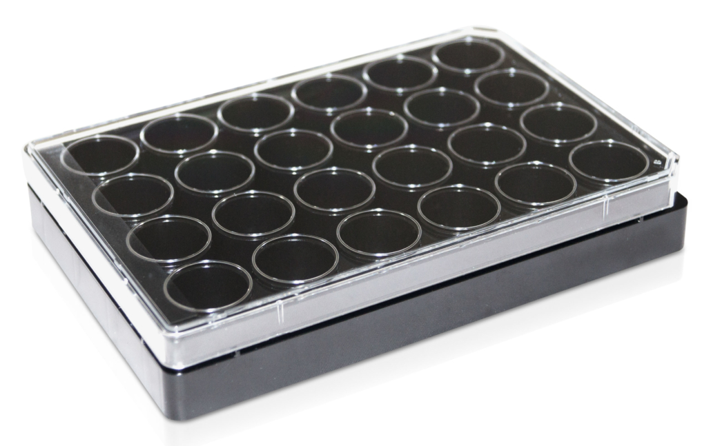
- Product Type: CytoSoft-Rigidity Plates
- Features: 2 kPa
- Packaging:1 x 24-well plate
- Features:Rigidity (elastic modules) 2 kPa
Cat.-Nr.: 5185-1EA

Description
CytoSoft®-Imaging products provide a tool to culture cells on PDMS substrates with various rigidity. This CytoSoft®-Imaging plate comes with 1 glass-bottom 24-well plate for high resolution imaging of cells on the selected stiffness.
The CytoSoft®-Imaging 24-well products have a defined elastic modulus (see certificate of analysis) with a #1.5 glass bottom attahced to a 24-well plate. The thickness of the silicone gel is uniform with a ~0.03 mm thick layer of silicone in each well. The silicone gels are activated and ready to bind to a purified ECM, such as PureCol® type I collagen (#5005) prior to cell addition.
The rigidity of the substrate to which cells adhere can have a profound effect on cell morphology and gene expression. CytoSoft® products provide a tool to culture cells on substrates with various rigidities covering a broad physiological range.
The CytoSoft® – Imaging plate is for high-resolution imaging where low autofluorescence and exceptional optical clarity are required. Plate consists of a 175 µm thin polycarbonate film-bottom plate bonded with a black polystyrene frame and includes a lid. The plates are sterilized using ozone and provided with 1 plate per package.
On the bottom of each well, there is a thin layer of specially formulated biocompatible silicone, whose elastic modulus (rigidity) is carefully measured and certified. The surfaces of the gels in CytoSoft® products are functionalized to form covalent bonds with amines on proteins. The chemical functionalization is stable and the reaction does not require a catalyst, facilitating coating of the gel surfaces with matrix proteins and plating cells.
The silicone substrates are optically clear and have a near zero auto-florescence. The layer of silicone in each well is firmly bonded to the bottom of the well. Unlike hydrogels (such as polyacrylamide gels), silicone gels are not susceptible to hydrolysis, do not dry or swell, are resilient and resistant to tearing or cracking, and their elastic moduli (rigidities) remain nearly unchanged during extended storage times.
CytoSoft®-Imaging products accommodate live cell staining using membrane and cell permeable dyes; fixation of cells using common techniques such as paraformaldexyde; and immunostaining of fixed cells.
CytoSoft coating reagents:
PureCol® (bovine collagen), Human Type III Collagen Solution, Human Type IV Collagen Solution, Human Fibronectin Solution, Human Vitronectin Solution
For an overview of our complete portfolio of CytoSoft® Cell Culture Ware and recommended coating matrices (click)
SUPPLIER:
Advanced BioMatrix
STATUS:
In Stock
SIZE:
1 pcs
 CytoSoft® Imaging, 24-well Plate, 8 kPa
You need to load content from reCAPTCHA to submit the form. Please note that doing so will share data with third-party providers.
More InformationYou are currently viewing a placeholder content from Turnstile. To access the actual content, click the button below. Please note that doing so will share data with third-party providers.
More InformationYou need to load content from reCAPTCHA to submit the form. Please note that doing so will share data with third-party providers.
More InformationYou are currently viewing a placeholder content from Facebook. To access the actual content, click the button below. Please note that doing so will share data with third-party providers.
More InformationYou are currently viewing a placeholder content from Instagram. To access the actual content, click the button below. Please note that doing so will share data with third-party providers.
More InformationYou are currently viewing a placeholder content from X. To access the actual content, click the button below. Please note that doing so will share data with third-party providers.
More Information