Matrix Proteins / Bioprinting
Advantages of Three-Dimensional (3D) Cell Culture Systems over traditional 2D Models
The behavior of cells cultured in a three-dimensional (3D) environment more accurately mirrors cellular responses observed in vivo. 3D cell culture systems or so-called 3D models (an in-vitro experimental set-up) better represent the complex microenvironment found within the body’s tissues. Extensive research indicates notable morphological and physiological differences between cells cultured in a 3D environment compared to those in a traditional two-dimensional (2D) culture setup.
Exploring the Dimensional Dynamics: Implications of 3D Culture in Cellular Responses and Disease Modeling
The pivotal aspect of 3D culture lies in its additional dimensionality, playing a crucial role in eliciting distinct cellular responses. This dimensionality not only influences the spatial organization of cell surface receptors engaged in interactions with neighboring cells but also imposes physical constraints on the cells themselves. These spatial and physical aspects in 3D cultures significantly impact signal transduction processes, bridging external stimuli to internal cellular responses. Therefore, a 3D model (i.e. skin model, lung model, atherosclerosis model) can mimic important steps for cardiovascular research, lung research, and cancer research. (cell adhesion, invasion of cells, transmigration of cells through an endothelial layer, impact on extracellular molecules)
Key Biomolecular Players in Shaping the Structural Integrity of 3D Cultures
Moreover, biomolecules such as collagen (atelocollagen and telocollagen), elastin, laminin, adhesion peptides, and adhesion molecules play a crucial role in the structure and function of 3D cultures. These proteins contribute to the formation and maintenance of the extracellular matrix, influencing cell adhesion and promoting a more realistic replication of the natural tissue environment. The stiffness of the 3D environment also contributes to the differentiation of cellular responses.
Exploring Substrate Rigidity: CytoSoft® Plates for Fine-Tuning Cell Culture Conditions
The rigidity of the substrate (stiffness) to which cells adhere can have a profound effect on cell morphology and gene expression. CytoSoft® plates provide a tool to culture cells on substrates with various rigidities covering a broad physiological range 0.2, 0.5, 2, 8, 16, 32, 64 kPa.
In addition, to analyze the impact of different stiffness of the gel itself, methacrylated matrix proteins can be used (PhotoCol®, PhotoGel®, and PhotoHA®).
Exploring 3D Bioprinting for Biomimetic Tissue Models
During the last years a new technique was established to build gels with a 3D printer (called bioprinting). These models include the same biomolecules such as collagen (atelocollagen and telocollagen), elastin, laminin, adhesion peptides, and adhesion molecules and gives us a new look inside this fantastic world.
-
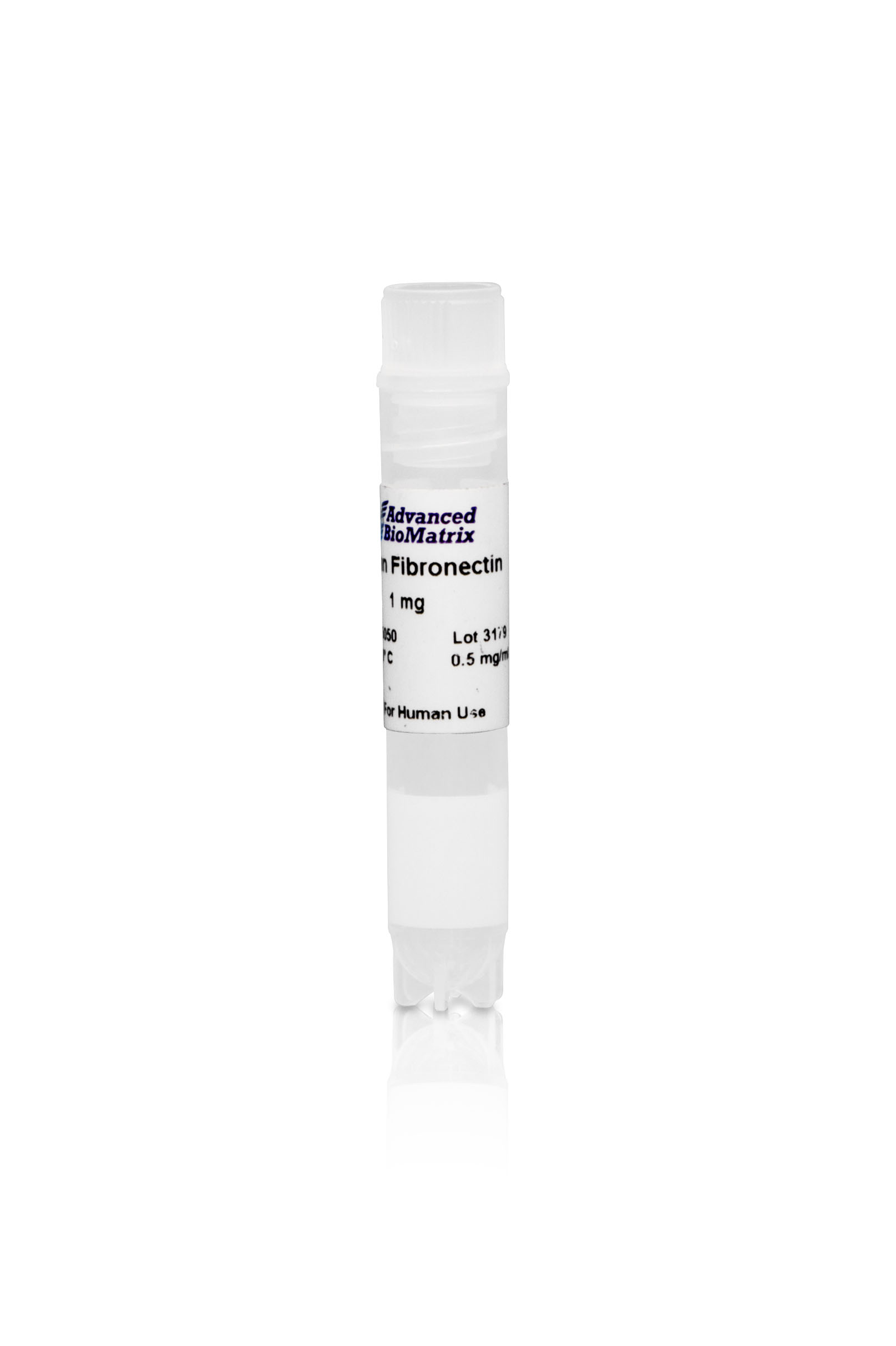
Human Fibronectin, Solution
Cat.-Nr: 5050-1MG
Fibronectin is a widely used broad range natural cell attachment factor. This product has been purified from human plasma where it is found as a... Read More
-
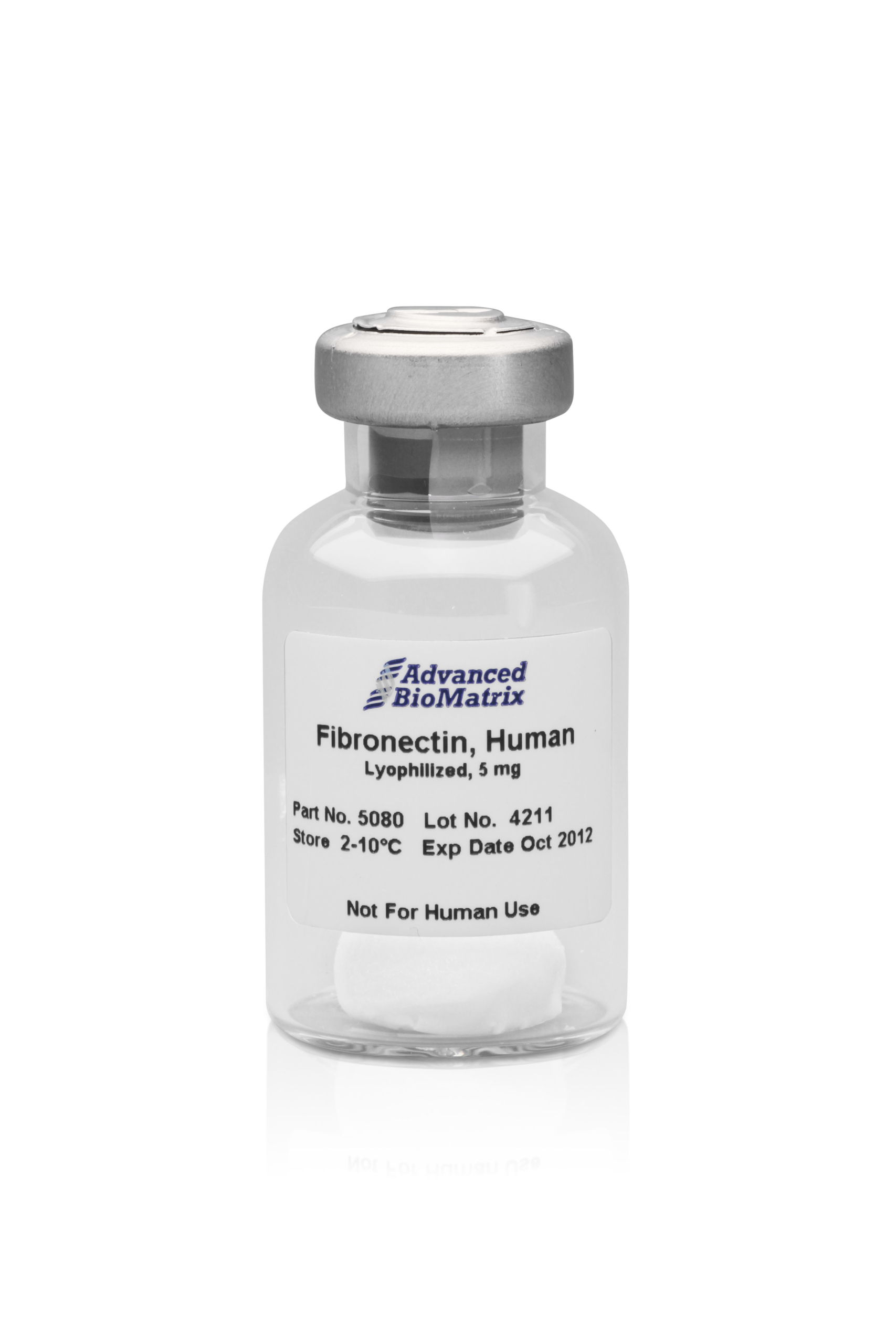
Fibronectin, lyoph. (human)
Cat.-Nr: 5080-5MG
Fibronectin is a widely used broad range natural cell attachment factor. This product has been purified from human plasma where it is found as a... Read More
-
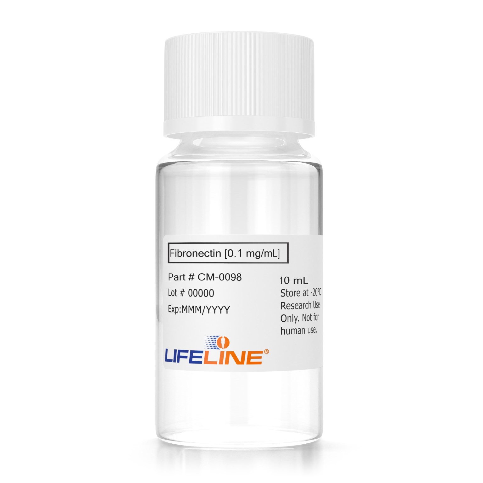
Fibronectin (0.1 mg/ml, 10 ml)
Cat.-Nr: CM-0098
Fibronectin is a purified protein commonly used as an extracellular matrix attachment factor. Lifeline® Fibronectin Solution, when used to pre-coat... Read More


