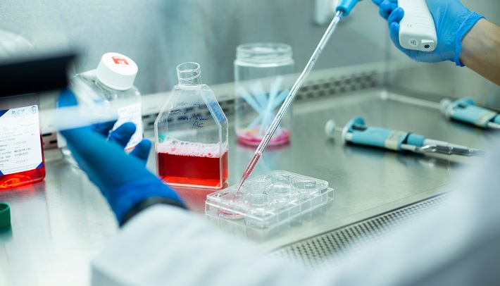
Advancing Hematologic Disease Research with 3D Bone Marrow Models
A major challenge in hematologic disease research is the development of model systems that accurately replicate human pathophysiology in vivo. Recent advancements in three-dimensional (3D) ex vivo modeling using human-induced pluripotent stem cell (iPSC)-derived bone marrow organoids provide a physiologically relevant approach to studying hematologic diseases.
Innovations in Bone Marrow Organoid Models
Recent studies have demonstrated that human iPSCs cultured in a hydrogel matrix enriched with human Collagen I (VitroCol, #5007-20mL) and human Collagen IV (#5022) support the formation of vascularized, myelopoietic bone marrow organoids. These matrices provide an optimized extracellular environment essential for the development of mesenchymal stromal cells, fibroblasts, endothelial cells, and hematopoietic cells, closely resembling the native bone marrow niche.[1-3]
Bone Marrow Organoids as a Platform for Hematologic Malignancies
Bone marrow organoids have been successfully used to support the engraftment, survival, and proliferation of hematopoietic stem and progenitor cells as well as primary cells from patients with myeloid and lymphoid blood cancers.[1]
Furthermore, it has been demonstrated that hematopoietic stem and progenitor cells (HSPCs) from patients with myelodysplastic syndromes (MDSs) efficiently engraft into bone marrow organoids, biomimicking the disease’s pathophysiology.[3]
The ability of these organoids to autonomously maintain hematopoiesis and sustain the marrow microenvironment underscores their potential as a transformative model for studying hematopoietic disorders and evaluating novel therapeutic strategies.
References
[1] Khan, A. et al. Cancer Discov (2023) 13 (2): 364–385.
[2] Olijnik, AA. et al. Nature Protocols (2024) 19: 2117–2146.
[3] Ren, K. et al. Blood Adv (2025) 9 (1): 54–65

